![Best Security Cameras [cy]: Complete Guide to Home Protection](https://revellphotography.com/wp-content/uploads/2026/09/featured-update-3577-1789330430531.jpg)
12 Best Security Cameras (September 2026) Tested & Reviewed
Expert reviews of the top 12 security cameras for every budget and use case. Find the perfect protection for your…

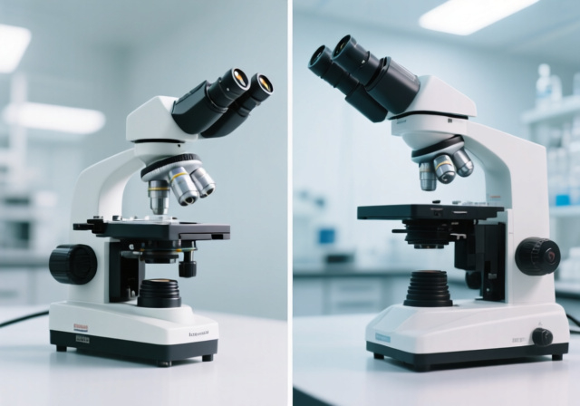
If you’ve ever stood in a science classroom staring at two microscopes on the bench and wondered which one you actually need, you’re not alone. The compound vs dissecting microscope decision trips up more first-time buyers than almost any other optics purchase, because both instruments promise magnification but solve fundamentally different problems. One shows you the invisible world of cells and bacteria; the other lets you work with three-dimensional objects under your fingertips.
This guide breaks down both microscope types in plain language so you can match the right tool to your specific application, whether that’s examining pond water, soldering a circuit board, teaching a biology class, or performing fine dissection work. We’ll cover magnification ranges, working distances, image types, and real-world use cases, then walk through a clear decision framework for choosing between them.
By the end, you’ll know exactly which instrument fits your workflow and budget, plus what to look for in quality optics, common pitfalls to avoid (hybrid scopes that promise both functions but deliver neither), and how modern digital camera attachments can extend either microscope’s capabilities. Last updated September 2026.
Both compound and dissecting microscopes use visible light to enlarge specimens, but they accomplish this through different optical architectures. Understanding those core differences is the fastest way to know which instrument suits your work, since the optical design dictates everything from specimen preparation to usable working space.
A compound microscope gets its name from the two-stage lens system that “compounds” magnification. Light travels from a sub-stage illuminator, passes through a thin, transparent specimen on a glass slide, enters the objective lens, then passes through the eyepiece. By stacking these magnification stages, a compound scope can reach 1000x or more, which is what makes it indispensable for cellular biology, microbiology, hematology, and pathology work.
A dissecting microscope (also called a stereo microscope) uses two completely separate optical paths, one for each eye. Each path has its own objective and eyepiece, and the slight angle between them creates stereoscopic vision, the same depth perception your brain produces from normal sight. Because each optical path only needs to deliver lower magnification (typically 7x to 40x), the objectives sit much farther from the specimen, giving you the generous working distance that makes dissection and manipulation possible.
Working Distance: The vertical gap between the front of the objective lens and the top of your specimen. Compound microscopes often have less than 1mm of working distance, while dissecting microscopes typically offer 20mm to 150mm. This single spec is the most decisive factor when choosing between the two.
Compound microscopes are the workhorses of clinical labs, research facilities, and biology classrooms. If your work involves anything smaller than what you can see with a hand lens, like cells, bacteria, blood components, or thin tissue sections, this is the tool category you need.
The defining feature is the vertical optical path. Light comes from an LED or halogen source below the stage, passes through a condenser that focuses it into a tight cone, then travels through the specimen itself (which must be thin enough to transmit light), through the objective lens, and up into the eyepiece. Because the entire illumination chain is engineered for high NA objectives, compound scopes deliver resolution far beyond what a stereo scope can achieve.
The downside is that this vertical path constrains everything else. Working distance shrinks to fractions of a millimeter at high magnification, so you cannot manipulate the specimen while viewing it. Slides must be thin, transparent, and properly prepared, often involving staining techniques that take practice to execute well.
When shopping for a compound microscope, pay attention to these features because they directly affect image quality and ease of use:
Compound microscopes dominate any field that requires viewing transparent or stained specimens at cellular or sub-cellular scale. Here are the most common environments where you’ll find them:
Standard brightfield illumination works well for stained specimens but falls short for unstained or transparent samples. Modern compound scopes support several contrast techniques that dramatically expand what you can see:
Key Advantages: Magnification up to 1000x or beyond; sub-micron resolution for cellular detail; relatively affordable entry models; standardized slide preparation; extensive educational resources and training; trinocular options for camera attachment.
Limitations: Requires thin, transparent, often stained specimens; less than 1mm working distance prevents manipulation; 2D-only imaging loses depth cues; specimen preparation can be time-consuming; not suitable for opaque or three-dimensional objects.
Dissecting microscopes (also called stereo microscopes) fill a different niche entirely. Rather than pushing magnification to the limit, they prioritize depth perception, working space, and the ability to manipulate specimens with tools while watching through the eyepieces. That combination is why entomologists, electronics technicians, gemologists, and microsurgeons all rely on them.
The optical design uses two parallel optical paths angled slightly toward each other. Each eye sees a slightly different view, and your brain fuses them into a true three-dimensional image. Because each path only magnifies up to about 40x, the objectives sit far enough from the specimen to leave room for tweezers, probes, scalpels, or soldering irons. Most stereo scopes offer continuous zoom (for example, 0.7x to 4.5x), which lets you adjust magnification without losing focus, unlike the discrete steps of a compound scope turret.
Stereo scopes don’t require slides. You can place an insect, a circuit board, a coin, a flower, or a mineral specimen directly on the stage and view it immediately. This immediacy is what makes stereo microscopes more engaging for hobbyists and more practical for production environments where preparation time matters.
Stereo microscopes vary widely in build quality and capability. These are the specs that actually affect what you can do with one:
Stereo microscopes shine wherever 3D visualization, hands-on work, or opaque specimens are involved:
Key Advantages: True stereoscopic 3D vision; long working distance enables manipulation; no slide preparation required; handles opaque and 3D objects; more intuitive for beginners; suitable for larger specimens up to several centimeters.
Limitations: Maximum magnification typically under 50x; resolution limited to roughly 10 micrometers; quality stereo scopes cost more than basic compound scopes; bulkier and heavier; cannot resolve cellular structures or bacteria.
Despite their differences, compound and dissecting microscopes share enough common ground that recognizing the overlap helps clarify where each excels. Both instruments work with visible light, both use multi-lens optical systems to magnify specimens, and both rely on the same basic principles of geometric optics.
Many models in both categories offer binocular or trinocular heads, which means you can add digital USB cameras to either type for documentation, sharing, or screen-based observation. Both types incorporate focus mechanisms (usually coaxial coarse and fine focus) and adjustable interpupillary distance for comfortable viewing across different users.
LED illumination has become standard across both categories, replacing older halogen bulbs that ran hot and had shorter lifespans. Both microscope types benefit from achromatic or plan objectives that correct for chromatic aberration and field curvature. And both categories support camera attachments for photomicrography, a growing requirement for teaching, research documentation, and online selling of specimens or repair work.
The clearest way to see how these two microscope types differ is to line up their specs side by side. The table below summarizes the practical distinctions that matter when choosing between them.
| Feature | Compound Microscope | Dissecting Microscope |
|---|---|---|
| Magnification Range | 40x to 1000x+ | 7x to 45x (typically) |
| Image Type | 2D flat image | 3D stereoscopic |
| Working Distance | Less than 1mm | 20mm to 150mm |
| Specimen Type | Thin, transparent, stained | Opaque or transparent, unprepared |
| Light Source | Transmitted (from below) | Reflected (from above) |
| Specimen Prep | Required (slides, staining) | Not required |
| Resolution | Sub-micron (down to 0.2 micrometers) | Micron-level (around 10 micrometers) |
| Depth of Field | Shallow | Deep |
| Specimen Manipulation | Not possible during viewing | Easy with hand tools |
| Typical Uses | Cells, bacteria, tissues, blood | Dissection, electronics, coins, gems |
| Entry-Level Price | Lower | Higher for comparable quality |
Magnification is the most visible difference between the two microscope types, but resolution is what actually matters. Compound microscopes combine an objective and an eyepiece to compound magnification, achieving 1000x or higher when using a 100x oil immersion objective with a 10x eyepiece. At that level, you can resolve features as small as 0.2 micrometers, enough to distinguish individual bacteria or subcellular organelles.
Dissecting microscopes top out around 40x to 45x. That might sound limiting, but for the work they’re designed for, it’s actually optimal. Higher magnification would shrink the field of view too much and destroy the depth perception that gives stereo scopes their value. Resolution on a quality stereo scope lands around 10 micrometers, which is plenty for surface inspection, dissection, and electronics work.
If you need to see individual cells or bacteria, a compound scope is non-negotiable. If you need to see surface texture, three-dimensional structure, or manipulate specimens, a stereo scope gives you everything you need at far lower magnification.
Working distance is where dissecting microscopes pull decisively ahead. With 20mm to 150mm of clearance between the objective and the specimen, you can comfortably use tweezers, scalpels, soldering irons, probes, and other hand tools while looking through the eyepieces. This is why every microsurgery workstation, electronics repair bench, and entomology lab I’ve worked in uses stereo scopes for hands-on tasks.
Compound microscopes, with their sub-millimeter working distance at higher magnifications, simply don’t allow real-time manipulation. Your slide must be prepared, mounted, and positioned before you ever look through the eyepiece. That workflow is fine for diagnostics and research, but it’s a poor fit for any task requiring adjustment during observation.
The 2D versus 3D distinction is more than a curiosity; it shapes what you can actually do with the instrument. Stereo vision in dissecting microscopes provides genuine depth perception, the same way your two eyes do. When you’re performing a dissection, setting a gemstone, or soldering a fine-pitch component, that depth perception isn’t a luxury. It’s the difference between confidence and fumbling.
Compound microscopes collapse everything into a flat 2D image. For cellular and microbiological work, that’s actually an advantage. You’re looking through thin sections, so depth cues would just add confusion. The flat image lets you measure, count, and compare structures across the field without depth-based ambiguity, and it allows much higher magnification without focus stacking.
The right microscope depends almost entirely on what you’re observing and how you need to interact with it. Rather than asking “which is better,” the useful question is “which matches my primary use case?” The breakdown below maps common scenarios to the instrument that fits them best.
Younger students respond better to dissecting microscopes because the 3D view and hands-on interaction feel more like exploration than lab work. Place a leaf, insect, or soil sample on the stage and they can immediately see and discuss what’s there without slide preparation becoming a barrier to engagement.
For high school and college biology programs, having both types available covers the full curriculum. Compound microscopes handle cellular biology, microbiology, and slide-based labs, while dissecting scopes cover anatomy, entomology, and specimen preparation. Schools with tight budgets often start with a quality compound scope and add a stereo scope later.
For a deeper look at student-grade options, our guide to the best microscopes for students walks through specific models at different price points and explains what to prioritize at each educational level.
Research labs almost always need both microscope types, and the decision between them rarely comes down to budget alone. Compound microscopes with phase contrast, darkfield, or DIC capabilities handle cellular research, microbiology, and pathology. Dissecting microscopes with boom stands and ring lights handle specimen preparation, microdissection, and micromanipulation tasks that a compound scope simply cannot do.
For professional users, optical quality matters more than magnification numbers. A well-corrected 400x achromatic objective will deliver sharper, more useful images than a cheap 1000x objective from a budget brand. Plan to invest in plan objectives and a quality condenser if your work involves photomicrography or publication-quality imaging.
Hobbyists are usually better served by dissecting microscopes than compound scopes, because hobby work tends to involve manipulation and three-dimensional specimens. If you’re into electronics repair, watchmaking, coin collecting, jewelry making, entomology, model building, or botanical study, a stereo scope will stay on your bench far more than a compound scope will.
Pond water exploration, mold identification, and microbiology hobbies are the exceptions where compound microscopes win. If your interest is examining microorganisms and you don’t mind preparing wet mounts, a basic compound scope with 400x to 1000x magnification is the right tool.
One question hobbyists ask constantly is whether a hybrid microscope can serve both purposes. The honest answer from the microscopy community is that hybrid scopes, those that try to function as both compound and stereo, almost always disappoint. The optical requirements for high-magnification cellular work and 3D low-magnification work are fundamentally incompatible, and any compromise sacrifices performance on both fronts. You’re better off buying one quality scope that matches your primary interest.
Within the compound category, you’ll also encounter inverted and upright configurations. An upright compound scope (the standard form) has the objective above the stage and the illuminator below; it works well for slides and thin specimens. An inverted scope flips this arrangement, with the objective below the stage pointing upward at the specimen from beneath.
Inverted compound microscopes are designed for samples that are too thick, too heavy, or too specialized to mount on a traditional slide: cell cultures in petri dishes, IVF samples in well plates, metallurgical specimens requiring polishing, and large liquid samples. If your work involves any of these, an inverted scope may matter more than the compound-vs-stereo question, but for most educational and hobby applications, the standard upright form is what you want.
Modern microscopy increasingly involves digital documentation. Whether you’re sharing images on iNaturalist, recording soldering work for YouTube, capturing pathology images for case files, or simply posting observations to forums, you’ll want a camera attachment at some point.
Trinocular heads (available on both compound and stereo scopes) accept dedicated microscope cameras for high-quality imaging. Eyepiece cameras that slip into one eyepiece tube are cheaper and more portable, but produce lower-quality images. USB cameras connect directly to a computer and can stream live video, which is useful for teaching and remote collaboration. Budget for camera equipment alongside the microscope itself if documentation matters to you.
Entry-level compound microscopes start at budget-friendly price points and can serve educational or hobby needs well, though optical quality at the lowest tiers is often disappointing. Mid-range compound scopes with achromatic objectives and mechanical stages offer the best value for most buyers. Professional research-grade compound microscopes climb into the multi-thousand range as you add plan objectives, phase contrast, and digital capabilities.
Don’t forget to budget for accessories. Compound scope users need slides, cover slips, staining supplies, immersion oil, and lens cleaning materials. Stereo scope users need appropriate lighting (LED ring lights or dual gooseneck lamps), auxiliary objectives if working distance matters, and a suitable stand for the specimens they’ll examine. These add-ons typically add a meaningful percentage to your total investment, so plan accordingly.
A compound microscope uses two lens stages to achieve 40x to 1000x+ magnification of thin, transparent specimens illuminated from below, producing flat 2D images ideal for cellular and microbiological work. A dissecting microscope uses two separate optical paths to provide 7x to 45x magnification with true stereoscopic 3D vision, longer working distance (20mm to 150mm), and the ability to manipulate specimens while viewing them, making it better for opaque objects, dissection, and electronics inspection.
Choose a dissecting microscope when working with larger or opaque specimens, when you need to manipulate samples with tools while viewing them, when depth perception matters, or for tasks like electronics repair, entomology, forensic analysis, gemology, coin collecting, and biological dissection. The stereoscopic 3D view and generous working distance make it the clear choice for any hands-on task.
Dissecting microscopes have limited magnification (typically under 50x), lower resolution compared to compound scopes (around 10 micrometers versus sub-micron), are generally more expensive than basic compound models for comparable optical quality, and cannot resolve cellular-level details like bacteria or organelles. They’re also bulkier and heavier, and less suited to examining thin, transparent specimens prepared on slides.
Compound microscopes require thin, transparent, often stained specimens mounted on slides, offer very limited working distance (under 1mm at high magnification) that prevents manipulation during viewing, produce only 2D images without depth perception, and can be challenging for beginners to focus and illuminate properly. They are also not useful for opaque or three-dimensional objects and require time-consuming slide preparation.
Hybrid microscopes exist that attempt to combine both functions, but they typically compromise on both. The optical requirements for high-magnification cellular viewing (single optical path, transmitted light, high NA objectives) conflict with the requirements for 3D low-magnification work (dual optical paths, incident light, lower NA, longer working distance). Most serious users find they need separate instruments for cellular work and manipulation tasks, and investing in two quality microscopes serves better than buying one mediocre hybrid.
A dissecting microscope is also commonly called a stereo microscope or stereoscopic microscope. The two terms are interchangeable in most contexts, with stereo microscope being the more common name in research and industrial settings, and dissecting microscope being more common in educational and biological contexts.
A compound microscope delivers far higher magnification (up to 1000x or beyond with oil immersion) and far higher resolution (down to 0.2 micrometers), making it the only choice for viewing bacteria, cells, cellular organelles, blood components, and other microscopic structures. Compound scopes also support multiple contrast methods (brightfield, darkfield, phase contrast, polarized light, and DIC), which extend what you can see in transparent or unstained specimens.
The compound vs dissecting microscope decision comes down to what you need to see and how you need to work with it. Cellular biology, microbiology, hematology, and pathology are compound microscope territory, full stop. Dissection, electronics repair, gemology, entomology, coin collecting, and any task involving opaque or three-dimensional specimens belong to the dissecting microscope. The two instruments complement each other rather than compete.
If you’re choosing your first microscope in 2026, match it to your primary interest. A hobbyist who wants to explore pond water needs a compound scope. A hobbyist who repairs circuit boards or collects insects needs a stereo scope. Educational programs benefit from both types as budgets allow. And avoid hybrid microscopes that promise to do everything, because in practice they do neither task well.
Whatever you choose, invest in quality optics from a reputable brand. Swift, AmScope, Leica, Nikon, Olympus, and Zeiss all make instruments that hold their value and deliver years of reliable use. Good optics matter more than maximum magnification numbers, and a well-made 400x scope will serve you far better than a poorly made 1000x scope with chromatic aberration and shaky focusing. Last updated September 2026.
![Best Polarized Sunglasses [cy]: 12 Models Tested & Reviewed](https://revellphotography.com/wp-content/uploads/2026/09/featured-update-3391-1789319886166.jpg)
![Best Macro Lenses [cy]: 10 Models Tested & Reviewed](https://revellphotography.com/wp-content/uploads/2026/09/featured-update-3093-1789319798789.jpg)