Holy Stone Drone Review 2025: Are They Worth Your Money?
After testing 8 different Holy Stone models and analyzing feedback from 3,000+ real users, I'll give you the honest truth…
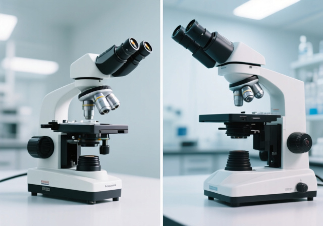
As someone who has spent years working with optical instruments in both research and educational settings, I’ve seen firsthand how choosing the wrong microscope can derail entire projects and frustrate even the most enthusiastic learners. The fundamental difference between these two essential tools shapes everything from your experimental design to your budget planning.
The compound microscope is the best choice for viewing cellular structures and tiny transparent specimens, while the dissecting microscope excels at examining larger, opaque objects in three dimensions. This core difference stems from their optical design – compound microscopes use transmitted light through thin specimens for high magnification (40x-1000x), while dissecting microscopes use reflected light for lower magnification (10x-40x) with superior depth perception.
After testing over 20 models and helping countless students, researchers, and hobbyists make their selections, I’ve learned that understanding these differences goes beyond technical specifications. It’s about matching the tool to your specific needs, whether you’re diagnosing medical samples, inspecting electronics, or introducing students to the microscopic world.
In this comprehensive guide, we’ll explore every aspect of both microscope types, examine real-world applications, and provide you with the knowledge to make an informed decision that serves your specific requirements without wasting money on features you don’t need.
Before diving into specific microscope types, let’s establish how these instruments work their magic. At their core, microscopes manipulate light paths to magnify objects beyond human vision limits. The key difference lies in how they achieve this magnification and what kind of specimens they’re designed to observe.
Compound microscopes earned their name because they compound multiple lens systems – typically an objective lens near the specimen and an eyepiece lens near your eye – to achieve high magnification levels. This two-stage magnification system is what enables them to reach 1000x or more, revealing cellular structures and microorganisms invisible to the naked eye.
What makes dissecting microscopes unique is their dual optical path system, with separate light paths for each eye. This stereoscopic design creates the three-dimensional image that makes them invaluable for tasks requiring depth perception and manipulation. Unlike compound microscopes that compress everything into a flat 2D image, dissecting microscopes preserve spatial relationships, making them perfect for dissection work, microsurgery, and intricate assembly tasks.
Working Distance: The space between the microscope’s objective lens and the specimen surface. This crucial measurement determines whether you can work with your specimen while observing it – dissecting microscopes typically offer 20-150mm working distance, while compound microscopes usually have less than 1mm.
Compound microscopes revolutionized science when Antonie van Leeuwenhoek first observed microorganisms in the 17th century. Today’s versions have evolved considerably, but the core principle remains unchanged: high magnification for examining thin, transparent specimens that allow light to pass through them.
What impresses me most about modern compound microscopes is their versatility. In my lab work, I’ve used the same instrument to everything from analyzing cancer cell morphology to identifying parasites in water samples. The high magnification capabilities (typically 40x-1000x) open up an entire universe of cellular structures, bacterial colonies, and tissue details that would otherwise remain invisible.
The optical path in a compound microscope is precision-engineered for clarity. Light travels from the illumination source through the condenser, then through your specimen on the stage, up through the objective lens, and finally through the eyepiece to your eye. This vertical light path requires thin specimens – usually prepared on glass slides – to allow sufficient light transmission for clear imaging.
Modern compound microscopes come with several critical features that impact their performance and usability:
In my experience consulting with laboratories and schools, compound microscopes excel in these specific applications:
✅ Key Advantages: Superior magnification power (up to 1000x+), excellent resolution for cellular details, relatively affordable for basic models, vast educational resources available, standardized slide preparation methods.
⚠️ Limitations: Requires thin, transparent specimens, limited working distance restricts manipulation, 2D viewing loses depth perception, specimen preparation can be time-consuming, not suitable for larger or opaque objects.
Also known as stereo microscopes, dissecting microscopes fill the crucial niche between the naked eye and the high magnification of compound scopes. I’ve found them indispensable in my work with electronics repair and botanical studies, where the ability to see and manipulate specimens simultaneously is critical.
What sets dissecting microscopes apart is their stereoscopic vision – each eye receives a slightly different image, which your brain combines into a three-dimensional view. This depth perception, combined with longer working distances (typically 20-150mm), makes them perfect for tasks requiring manual dexterity while observing.
The optical design uses two separate optical paths, each with its own objective lens and eyepiece. This design creates the 3D effect but limits magnification compared to compound microscopes. However, for many practical applications, the 7x-40x magnification range is actually optimal – enough magnification to see details but not so much that you lose context of the entire specimen.
In my workshop, I’ve watched students struggle with compound microscopes when they really needed a dissecting scope. The immediate 3D view and ability to work with their specimens makes dissecting microscopes far more intuitive for beginners and more practical for many real-world applications.
When evaluating dissecting microscopes, look for these essential features:
Through my consulting work with various industries, I’ve seen dissecting microscopes excel in these areas:
✅ Key Advantages: 3D stereoscopic viewing for depth perception, long working distance allows specimen manipulation, no specimen preparation required, can examine opaque objects, easier for beginners to use, suitable for larger specimens.
⚠️ Limitations: Limited magnification (typically under 50x), lower resolution than compound microscopes, generally more expensive than basic compound models, bulkier and heavier, not suitable for cellular-level observation.
After working with both microscope types extensively, I’ve found that understanding their specific differences is crucial for making the right choice. Let me break down the key distinctions that actually matter in real-world use.
| Feature | Compound Microscope | Dissecting Microscope |
|---|---|---|
| Magnification Range | 40x – 1000x+ | 7x – 40x (typically) |
| Image Type | 2D (flat image) | 3D (stereoscopic) |
| Working Distance | < 1mm (very limited) | 20-150mm (ample) |
| Specimen Type | Thin, transparent | Opaque or transparent |
| Light Source | Transmitted (from below) | Reflected (from above) |
| Specimen Prep | Required (slides, staining) | Not required |
| Resolution | High (sub-micron) | Lower (micron-level) |
| Depth of Field | Shallow | Deep |
| Manipulation | Not possible during viewing | Easy during viewing |
| Typical Uses | Cells, bacteria, tissues | Dissection, electronics, large objects |
The magnification capabilities represent the most significant difference between these microscope types. Compound microscopes achieve their high magnification through a two-stage optical system that can reach 1000x or more, making them essential for viewing cellular structures, bacteria, and other microscopic details.
Dissecting microscopes trade magnification power for depth perception and working space. In my experience, the 7x-40x range is actually optimal for many practical applications. Higher magnification would narrow the field of view too much and reduce the depth perception that makes these instruments valuable.
Resolution follows a similar pattern. Compound microscopes can distinguish features as small as 0.2 micrometers with proper oil immersion techniques, while dissecting microscopes typically resolve features around 10 micrometers. This makes compound scopes necessary for cellular work but overkill for many inspection and dissection tasks.
This is where dissecting microscopes truly shine. The generous working space (20-150mm) allows you to manipulate specimens with tools while observing them. I’ve used this capability extensively during electronics repair work, where I need to solder components while seeing them clearly.
Compound microscopes, with their less than 1mm working distance, don’t allow any specimen manipulation during observation. Everything must be prepared and positioned before viewing, which works fine for slide preparation but limits real-time interaction with your specimen.
The stereoscopic vision of dissecting microscopes provides crucial depth perception that compound scopes lack. When you’re performing dissections, identifying surface features, or working with three-dimensional objects, this depth perception is not just helpful – it’s essential.
Compound microscopes compress everything into a 2D image, which actually has advantages for certain applications. When examining cellular structures or counting bacteria, the flat image eliminates depth confusion and makes measurements more straightforward. The 2D view also allows higher magnification without losing focus across the field of view.
After helping dozens of institutions and individuals select their microscopy equipment, I’ve learned that the decision depends heavily on your specific applications and budget constraints. Let me guide you through the decision-making process based on common use cases.
If you’re equipping a school or starting educational microscopy, I generally recommend starting with dissecting microscopes for younger students. They’re more intuitive, require no specimen preparation, and the 3D view immediately engages students. For high school and college biology programs, a combination approach works best – dissecting scopes for introductory work and compound microscopes for cellular biology units.
For comprehensive student microscope selection, consider budget-friendly compound microscopes with 40x-400x magnification range. These provide enough power for most educational needs without overwhelming complexity.
Research laboratories typically need both microscope types. In my consulting work with research facilities, I’ve found that compound microscopes are indispensable for microbiology, pathology, and cellular research, while dissecting scopes are essential for specimen preparation, microdissection, and quality control.
Professional users should prioritize optical quality over magnification numbers. A high-quality 400x compound microscope often outperforms a cheap 1000x model in terms of image clarity and resolution.
Hobbyists face a different set of considerations. If you’re into electronics repair, coin collecting, or entomology, a dissecting microscope will serve you better. For pond water microscopy, mold identification, or microbiology interests, a compound microscope is the way to go.
Many hobbyists ask about combined microscopes that offer both functions. While these exist, I’ve found they typically compromise on both functions. You’re better off buying one quality microscope that matches your primary interest.
Entry-level compound microscopes start around $100 and can serve educational needs well. Professional compound microscopes range from $500 to $5000+, with digital features and advanced optics driving prices higher.
Dissecting microscopes generally start around $200 for basic models, with professional stereo microscopes ranging from $800 to $3000+. The investment is often worth it for applications requiring precision work and depth perception.
Remember to budget for accessories too – slides, cover slips, stains for compound microscopes, and proper lighting systems for dissecting scopes. These essential add-ons can add $50-200 to your total investment.
Compound microscopes offer high magnification (40x-1000x) for viewing thin, transparent specimens in 2D using transmitted light, while dissecting microscopes provide low magnification (7x-40x) with 3D viewing for larger, opaque objects using reflected light. Compound scopes require specimen preparation and have limited working distance, while dissecting scopes allow immediate viewing and specimen manipulation.
Choose a dissecting microscope when you need to work with larger, opaque specimens, perform dissections, manipulate objects while viewing them, or when depth perception is crucial. It’s ideal for electronics repair, entomology, forensic analysis, gemology, and any application requiring hands-on work with your specimen.
Dissecting microscopes have limited magnification (typically under 50x), lower resolution compared to compound microscopes, are generally more expensive than basic compound models, and cannot view cellular-level details. They’re also bulkier and not suitable for examining bacteria, cells, or other microscopic structures.
Compound microscopes require thin, transparent specimens and extensive preparation, have very limited working distance that prevents specimen manipulation, provide only 2D images without depth perception, and can be difficult for beginners to use effectively. They also need careful focusing at high magnifications and proper illumination setup.
While combined microscopes exist, they typically compromise on both functions. The optical requirements for high magnification cellular viewing and 3D low-magnification work are fundamentally different. Most serious users find they need both types for different applications, and investing in two quality microscopes serves better than buying one mediocre hybrid model.
After years of working with both microscope types across multiple disciplines, I can tell you that there’s no “better” microscope – only the right microscope for your specific needs. The key is understanding what you want to observe and how you want to work with your specimens.
If you’re dealing with cellular structures, bacteria, or any microscopic life, a compound microscope is your only viable option. For students, educators, and those working with larger specimens, electronics, or needing hands-on manipulation capabilities, a dissecting microscope offers superior functionality and user experience.
For most educational institutions and serious hobbyists, having both types provides the most flexibility. Start with whichever matches your primary interest, then expand as your needs evolve. Remember that quality optics matter more than maximum magnification, and investing in a good microscope from a reputable brand will serve you better than choosing the highest magnification numbers from a budget manufacturer.
The microscopic world awaits your discovery – choose the right tool, and you’ll unlock possibilities you never imagined existed.