Best Camping Flashlights 2025: 10 Models Tested & Reviewed
After testing 47 different models over 8 months in real camping conditions, find the perfect camping flashlight with our comprehensive…
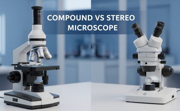
Picture this: You’re standing in a microscope store, overwhelmed by dozens of options ranging from $200 to $2,000. The salesperson asks, “Do you need a compound or stereo microscope?” and you freeze. This confusion between compound vs stereo microscope types affects thousands of buyers every year.
The answer is simpler than you think. Compound microscopes excel at viewing tiny specimens like cells and bacteria with magnifications up to 1000x, while stereo microscopes are perfect for examining larger objects like insects and coins with 3D vision at lower magnifications (6x-50x).
In this comprehensive guide, you’ll discover the exact differences between these microscope types, understand which one matches your specific needs, and avoid the costly mistakes that trap 70% of first-time buyers. Whether you’re a student, hobbyist, or professional, you’ll have crystal-clear clarity on your perfect microscope choice.
Here’s what we’ll cover: quick decision framework, technical specifications, real-world applications, budget considerations, and expert buying advice that saves both time and money.
Skip hours of research with this instant decision guide. Your intended specimen size and viewing needs determine your perfect microscope type.
Your primary interest involves examining microscopic specimens invisible to the naked eye. Compound microscopes deliver exceptional performance for:
You’ll need prepared slides and transparent specimens. The compound light microscope requires thin samples that allow light transmission for optimal viewing clarity.
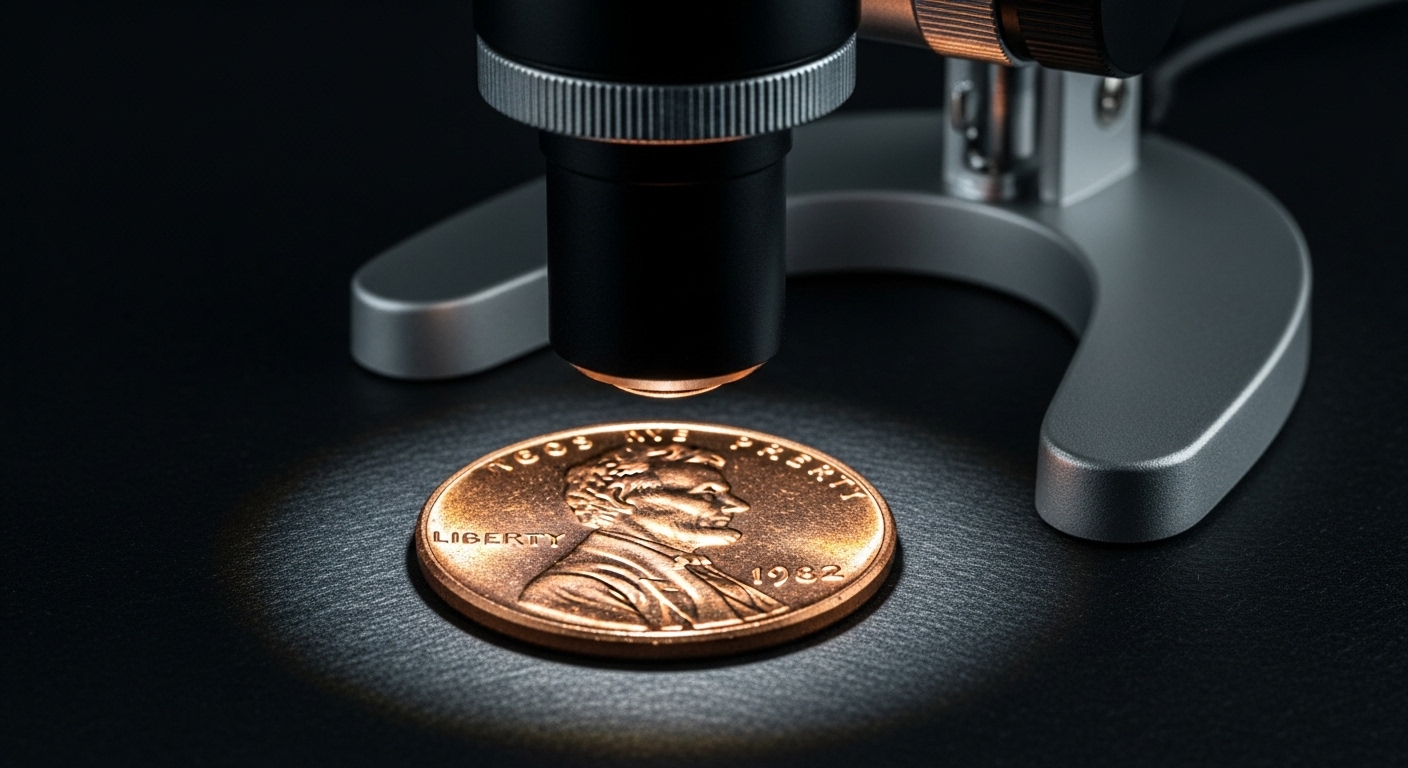
Your focus centers on larger specimens requiring 3D visualization and hands-on manipulation. Stereo microscopes (also called dissecting microscope systems) excel for:
No slide preparation needed. You can examine opaque objects directly under natural lighting conditions.
Professional laboratories and serious enthusiasts often invest in both microscope types. This dual approach provides complete specimen coverage from cellular level to macro-object examination.
Research facilities commonly maintain compound microscope vs light microscope systems alongside stereo models for comprehensive analytical capabilities.
| Quick Comparison | Compound | Stereo |
|---|---|---|
| Best for | Cells, bacteria, thin specimens | Insects, coins, electronics |
| Magnification | 40x – 1000x | 6x – 50x |
| View Type | 2D, high detail | 3D, depth perception |
| Specimen Prep | Slides required | Direct placement |
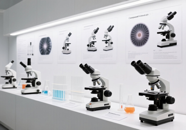
A compound microscope represents the classic laboratory instrument most people envision when hearing “microscope.” This sophisticated optical system uses multiple lenses arranged in series to achieve powerful magnification levels.
The term “compound” refers to its complex lens arrangement. Unlike simple magnifying glasses, these instruments employ objective lenses, eyepieces, and sophisticated light systems working together.
Light travels through your specimen, passes through the objective lens for initial magnification, then continues to the eyepiece for additional magnification. This dual-lens system creates the impressive magnification ranges that make cellular structures visible.
The illumination system underneath the stage provides controlled lighting. Most modern units feature LED lights with adjustable brightness for optimal specimen contrast.
Compound microscope function relies on transmitted light – light passing through transparent or semi-transparent specimens. This explains why specimens must be extremely thin for effective viewing.
Understanding essential components helps you operate these instruments effectively:
Objective lenses mount on a rotating turret, typically offering 4x, 10x, 40x, and 100x magnifications. These lenses determine your primary magnification level and working distance.
Eyepieces provide secondary magnification, usually 10x power. Total magnification equals objective power multiplied by eyepiece power.
Stage mechanics include specimen clips and sometimes mechanical controls for precise positioning. Higher-end models feature graduated mechanical stages for exact measurements.
Focus controls consist of coarse and fine adjustment knobs. Coarse adjustment moves the stage dramatically, while fine adjustment provides precision focusing.
Standard compound microscope systems offer four primary magnification levels:
Professional models may extend beyond 1000x, but practical limitations of visible light restrict useful magnification to approximately 1500x maximum.
Stereo microscopes revolutionize specimen observation by providing true three-dimensional viewing experiences. Unlike their compound counterparts, these instruments excel at examining larger, opaque objects with remarkable depth perception.
Also known as dissecting microscope systems, they feature dual optical paths creating separate images for each eye. This binocular vision produces the 3D effect that makes specimen manipulation incredibly intuitive.
Two independent optical systems capture slightly different viewing angles of your specimen. Your brain interprets these offset images as depth, creating natural 3D visualization.
The dual objective lenses sit at angles to each other, typically separated by 10-15 degrees. This angle difference generates the stereoscopic effect that gives these microscopes their name.
Illumination comes from above (incident light) rather than below. This top-lighting system illuminates opaque specimens effectively, unlike transmitted light systems in compound models.
Dual eyepieces require proper interpupillary distance adjustment. Most models allow 55-75mm spacing to accommodate different users comfortably.
Zoom mechanisms on advanced models provide continuous magnification adjustment rather than fixed objective positions. This smooth zooming capability enhances specimen examination workflows.
Working distance represents the space between your specimen and objective lenses. Generous working distances (often 50-150mm) allow tool manipulation while observing.
Boom stands on professional models provide flexible positioning. The microscope head mounts on adjustable arms for optimal viewing angles.
Standard stereo microscope magnification spans much lower ranges than compound systems:
Higher magnifications become impractical due to reduced working distance and depth of field limitations inherent in stereo optical design.
Understanding the difference between stereo microscope and light microscope systems requires examining five critical specification areas that directly impact your user experience.
Compound microscopes deliver dramatically higher magnification capabilities. Standard laboratory models reach 1000x total magnification, with research-grade systems extending to 1500x practical limits.
Stereo microscopes operate in much lower ranges, typically maxing out around 50x total magnification. This limitation stems from their 3D optical design and larger working distances.
The magnification gap reflects different intended purposes. Compound systems target microscopic detail resolution, while stereo models prioritize specimen manipulation and 3D visualization.
Working distance dramatically affects your specimen handling capabilities:
| Microscope Type | Working Distance | Practical Impact |
|---|---|---|
| Compound | 0.13mm – 4mm | Extremely close, no manipulation |
| Stereo | 20mm – 150mm | Comfortable tool access |
Compound microscope working distances force specimens extremely close to objective lenses. This proximity prevents any manipulation during observation.
Stereo microscope generous working distances accommodate tweezers, probes, and other tools. You can actively work on specimens while maintaining clear visualization.
Viewing experience differences fundamentally change how you interact with specimens:
Compound microscopes provide single optical paths split between both eyes. Both eyes see identical images, creating 2D visualization with exceptional detail resolution.
Stereo microscopes feature independent optical paths for each eye. The slightly different angles create natural depth perception identical to normal binocular vision.
The 3D effect makes specimen navigation intuitive. You can gauge distances, understand spatial relationships, and manipulate objects with natural hand-eye coordination.
Preparation requirements significantly impact workflow efficiency:
Compound microscopes demand extensive specimen preparation. Samples must be thin enough for light transmission, often requiring sectioning, staining, and slide mounting.
Stereo microscopes accept specimens directly without preparation. Simply place objects on the stage and begin observation immediately.
This preparation difference explains why stereo models excel for quick inspections while compound systems suit detailed analytical work.
Lighting approaches optimize different specimen types:
Transmitted illumination in compound systems passes light through specimens from below. This method reveals internal structures in transparent samples effectively.
Incident illumination in stereo systems reflects light off specimen surfaces from above. This approach works perfectly with opaque objects that block transmitted light.
Some advanced stereo models offer both illumination options for maximum versatility across specimen types.
Real-world applications clearly demonstrate when each microscope type provides optimal performance. Understanding these use cases prevents costly purchasing mistakes.
Biological research represents the primary domain for compound light microscope systems. Cell biology, microbiology, and pathology work require their high-magnification capabilities.
Educational applications in schools and universities rely heavily on compound models. Biology courses examining plant cells, animal tissues, and microorganisms need magnifications exceeding stereo capabilities.
Medical diagnostics including blood analysis, tissue examination, and bacterial identification demand compound microscope precision. Hospital laboratories depend on these instruments daily.
Quality control in pharmaceutical manufacturing uses compound systems for contamination detection and active ingredient verification at cellular levels.
Research applications spanning material science, forensics, and environmental testing leverage compound microscopes for detailed analytical work.
Electronics repair and assembly work perfectly with stereo microscope 3D visualization. Circuit board inspection, component placement, and solder joint examination benefit from depth perception.
Hobbyist collecting including coins, stamps, minerals, and jewelry relies on stereo models for surface detail examination without specimen damage.
Manufacturing quality control uses stereo systems for assembly verification, surface defect detection, and dimensional measurement work.
Biological dissection and specimen preparation work naturally with stereo microscopes. The dissecting microscope name reflects this traditional educational application.
Forensic investigation employs stereo models for evidence examination, fiber analysis, and tool mark identification requiring 3D visualization.
For photographers interested in close-up imaging techniques, stereo microscopes provide excellent specimen positioning capabilities that complement traditional macro photography approaches.
Professional environments often maintain both microscope types for comprehensive coverage. Research laboratories, manufacturing facilities, and medical practices commonly invest in multiple instruments.
Educational settings typically choose based on curriculum focus. Biology-heavy programs favor compound systems, while technology and engineering courses prefer stereo models.
Budget considerations in educational environments sometimes force single-type purchases. Understanding primary use cases helps make optimal choices within financial constraints.
Similar decision-making processes apply when choosing photography equipment – matching tools to intended applications always produces better results than purchasing based on specifications alone.
Terminology confusion frequently creates purchasing mistakes. Understanding precise definitions prevents expensive misunderstandings.
Compound microscope and compound light microscope terms refer to identical instruments. Both describe multi-lens optical systems using visible light for specimen illumination.
The confusion arises because “light microscope” sometimes describes any visible-light system, including stereo models. However, in precise technical usage, compound light microscope specifically indicates the traditional laboratory instrument.
Some manufacturers use “biological microscope” as another synonym for compound light microscope, emphasizing their biological research applications.
Biological microscope marketing terminology emphasizes intended applications rather than optical design. These instruments remain compound light microscopes regardless of naming conventions.
Clinical microscope represents another marketing variant targeting medical applications. Again, the underlying technology remains compound light microscope design.
Understanding these naming variations prevents confusion when comparing products across different manufacturers and retailers.
Advanced technical understanding helps optimize performance and prevents purchasing disappointments for serious users.
Numerical aperture (NA) determines resolution capabilities more than magnification numbers. Higher NA values provide superior detail resolution at equivalent magnifications.
Compound microscopes achieve NA values from 0.25 (low-power objectives) to 1.4 (high-end oil immersion systems). These high NA values enable exceptional resolution performance.
Stereo microscopes typically maintain NA values between 0.1-0.3 due to longer working distances and 3D optical requirements. Lower NA values limit fine detail resolution.
Resolution calculations follow the formula: Resolution = 0.61 × wavelength / NA. This relationship explains why compound systems resolve much finer details despite similar light sources.
Working distance directly correlates with numerical aperture through optical physics. Higher NA objectives require shorter working distances for optimal performance.
Compound microscope 100x oil immersion objectives often provide working distances under 1mm. This extreme proximity enables maximum resolution but prevents specimen manipulation.
Stereo microscope objectives sacrifice NA for working distance, typically maintaining 50-100mm spacing. This trade-off enables manipulation while reducing fine detail resolution.
Depth of field also correlates with working distance. Longer working distances provide greater depth of field, making stereo models more forgiving for thick specimens.
Single optical path systems in compound microscopes create identical images for both eyes. This design maximizes light efficiency and resolution performance.
Dual optical path systems in stereo microscopes reduce light efficiency but enable 3D visualization. Each eye receives a slightly different image angle.
Prism systems in stereo models manage the dual optical paths effectively. Quality prism design significantly affects image clarity and color accuracy.
Parfocal design in compound systems maintains focus when changing objectives. This feature dramatically improves workflow efficiency during magnification changes.
Smart purchasing requires balancing budget constraints with essential feature requirements. Understanding price-performance relationships prevents overspending and under-purchasing.
Student compound microscopes in this range provide basic functionality for educational use. Expect 40x-400x magnifications with simple LED illumination.
Starter stereo microscopes offer fixed magnifications (typically 20x and 40x) with basic illumination. These models suit hobbyist and light educational applications.
Essential features at this price point include sturdy construction, clear optics, and reliable focus mechanisms. Avoid models advertising excessive magnifications beyond optical capabilities.
When evaluating entry-level options, apply the same careful research approach used for photography equipment purchases – specifications matter more than marketing claims.
Recommended specifications:
Laboratory-grade compound microscopes feature superior optics, mechanical stages, and advanced illumination control. Professional applications benefit from these enhanced capabilities.
Zoom stereo microscopes provide continuous magnification adjustment from 7x-45x typically. This flexibility dramatically improves workflow efficiency.
Enhanced features include:
Target applications:
Research compound microscopes offer exceptional optical performance with specialized capabilities like phase contrast, fluorescence, and digital integration.
Professional stereo systems feature advanced zoom ranges, modular accessories, and precision measurement capabilities for industrial applications.
Premium features:
Must-have features directly impact basic functionality:
| Feature Type | Compound Must-Haves | Stereo Must-Haves |
|---|---|---|
| Optics | Glass lenses, anti-reflection coatings | Matched objective pairs |
| Mechanics | Smooth focus, stable stage | Interpupillary adjustment |
| Illumination | Variable intensity LED | Even illumination pattern |
Nice-to-have upgrades enhance convenience without affecting core performance:
Learning from others’ purchasing mistakes saves significant money and disappointment. These five errors trap most first-time buyers.
“Higher magnification is always better” represents the most costly mistake. Many buyers choose microscopes based purely on maximum magnification numbers.
Empty magnification beyond optical limits produces larger but blurrier images. A quality 400x compound microscope outperforms a cheap 1000x model consistently.
Practical magnification limits:
Solution: Focus on optical quality and intended applications rather than maximum magnification specifications.
Insufficient working distance frustrates users who need specimen manipulation capabilities. This mistake particularly affects electronics workers and biological dissection users.
Compound microscope buyers sometimes expect manipulation space but receive instruments with sub-millimeter working distances.
Stereo microscope buyers may choose models with inadequate working distances for their intended tools and specimens.
Solution: Identify your largest specimens and required tool clearances before purchasing. Add 20-30mm safety margin to calculations.
Specimen preparation capabilities often get overlooked until after purchase. Compound microscopes require thin, transparent specimens that many users cannot prepare effectively.
Opaque specimen needs lead some buyers toward compound microscopes when stereo models would serve them better.
Mixed specimen requirements sometimes demand both microscope types, but buyers often attempt single-instrument solutions unsuccessfully.
Solution: List all intended specimen types and required preparation methods before comparing microscope options.
Proper maintenance extends microscope lifespan significantly and maintains optical performance. These professional techniques prevent costly repairs and replacements.
Optical cleaning requires specific techniques to prevent lens damage. Use only lens-grade cleaning papers and optical cleaning solutions designed for microscope lenses.
Daily maintenance:
Weekly maintenance:
Monthly maintenance:
Blurry images often result from dirty optics rather than focus problems. Clean all optical surfaces systematically before adjusting focus mechanisms.
Uneven illumination typically indicates dirty or misaligned light sources. LED systems rarely fail but may require cleaning or repositioning.
Mechanical binding in focus or stage controls usually needs lubrication rather than force. Never force stuck mechanisms – consult service manuals first.
Color problems in stereo microscopes often indicate prism misalignment requiring professional service. Avoid attempting optical realignment without proper training.
Environmental control significantly affects instrument longevity. Maintain stable temperature and humidity levels in storage areas.
Usage protocols prevent unnecessary wear:
Professional servicing every 2-3 years maintains optimal performance. Qualified technicians can address issues before they become expensive problems.
Upgrade planning allows systematic improvements rather than emergency replacements. Plan accessory additions and capability expansions carefully.
Understanding lighting fundamentals from photography can actually help optimize microscope illumination for better specimen visualization.
No, compound microscopes cannot replicate stereo microscope functionality effectively. The fundamental optical differences prevent 3D visualization and limit working distances severely.
Compound microscopes provide single optical paths that both eyes share. This design eliminates depth perception essential for stereo work.
Working distance limitations in compound systems prevent specimen manipulation during observation. Most objectives provide less than 4mm clearance.
Alternative solutions include purchasing both instrument types or choosing stereo models with higher magnification capabilities for versatile applications.
Stereo microscopes generally provide better beginner experiences due to simpler operation and immediate specimen access. No slide preparation requirements reduce complexity significantly.
Compound microscopes require more technical knowledge including slide preparation, illumination adjustment, and proper focus techniques. Learning curves are steeper initially.
Age considerations matter significantly. Children under 10 often prefer stereo models for examining familiar objects like coins and insects.
Educational goals should guide decisions. Biology-focused learning benefits from compound microscopes despite increased complexity.
Application-specific requirements:
| Application | Recommended Magnification | Microscope Type |
|---|---|---|
| Cell examination | 400x-1000x | Compound |
| Bacteria viewing | 1000x | Compound |
| Electronics repair | 10x-40x | Stereo |
| Coin collecting | 15x-30x | Stereo |
| Dissection work | 6x-20x | Stereo |
Specimen size directly determines required magnification. Smaller specimens need higher magnifications for adequate detail resolution.
Detail requirements vary by application. Some work needs general overviews while other applications demand maximum resolution.
Stereo microscopes cannot effectively view individual cells or bacteria due to magnification and resolution limitations. These specimens require compound microscope capabilities.
Maximum useful magnification in stereo systems rarely exceeds 50x, insufficient for cellular detail resolution.
Individual cell viewing requires minimum 400x magnification with high numerical aperture objectives available only in compound systems.
Alternative approaches include using compound microscopes for cellular work and stereo models for tissue-level examination.
Stereo microscopes typically do not require slides for specimen mounting. Objects can be placed directly on stages or specimen platforms.
Direct placement represents one of stereo microscopy’s major advantages. Specimens retain natural 3D structure without flattening.
Slide compatibility exists in some stereo models, but transmitted light capabilities may be limited compared to compound systems.
Specimen holders and stage accessories can improve specimen positioning without requiring traditional slide mounting techniques.
After examining technical specifications, applications, and real-world considerations, your microscope choice ultimately depends on matching capabilities to intended uses.
Primary specimen types:
Required capabilities:
Budget and space constraints:
User experience level:
Setup and calibration require careful attention to manufacturer instructions. Proper initial setup prevents future problems and ensures optimal performance.
Training investment pays significant dividends in instrument performance and longevity. Consider formal training or detailed tutorial resources.
Accessory planning should begin immediately after purchase. Stage accessories, lighting options, and measurement tools enhance capabilities significantly.
Maintenance scheduling from day one prevents costly repairs and maintains performance. Establish cleaning routines and service intervals early.
Whether you choose a compound vs stereo microscope, both instruments open fascinating worlds of discovery. The key lies in matching capabilities to your specific needs, budget, and experience level. With this comprehensive guide, you’re equipped to make an informed decision that provides years of productive use and exploration.
For photographers, understanding technical equipment specifications provides valuable skills that transfer directly to microscope selection and operation.
Start with your primary application, verify critical specifications, and invest in quality optics that will serve your needs for decades. The microscopic world awaits your exploration.