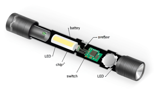
How Does a Flashlight Work: Complete Guide 2026
Discover how flashlights convert electricity into light through simple circuits, LED technology, and the components that make modern portable lighting…
Long before a single lens was ground into a brass tube, humans were already curious about the tiny world just past the edge of sight. From the sand grains of a desert to a single drop of pond water, the instinct to look closer is ancient. Microscopes turned that instinct into a science, and the microscope facts we have collected over four centuries tell a story of stubborn curiosity, clever glass-blowing, and a few happy accidents along the way.
This guide walks through the history, the engineering, and the modern applications of microscopy. You will meet the lensmakers, the biologists, and the physicists who pushed magnification past the visible, and you will learn why a single drop of water under a Leeuwenhoek-style scope still thrills hobbyists today. Whether you are a student, a teacher, or simply someone who wants to understand how scientists see the invisible, these microscope facts will give you a working mental model of one of humanity’s most important inventions.
A microscope is a scientific instrument that uses one or more lenses to produce enlarged images of objects too small to resolve with the naked eye. The word itself is a Latin-Greek hybrid coined in 1625 by Giovanni Faber, a member of the Accademia dei Lincei, who paired the Greek mikros (small) with skopein (to look or to examine). The Latin form, microscopium, gave us the modern English spelling we use today.
Modern instruments can magnify from roughly 10x on a basic stereo model all the way past 10 million times on a transmission electron microscope. That range covers everything from the weave of a single thread to the lattice structure of a crystal. The earliest handheld scopes offered perhaps 9x magnification, yet they were enough to ignite a chain of discoveries that defined modern biology, medicine, and materials science.
At its core, a microscope is a wave-bending tool. Whether the wave is a beam of visible light or a stream of accelerated electrons, the same trick is at work: the instrument shapes the wave so that detail hidden by the limits of human vision is enlarged enough to be seen, photographed, or measured.
Etymology box: Why is it called a microscope? The term comes from the Greek mikros (“small”) and skopein (“to look at”). Italian scholar Giovanni Faber coined the Latin compound microscopium in 1625, the same year he proposed the name “telescope” for Galileo’s far-seeing instrument. The two words were born as a matched pair.
The story of the microscope is older than most people realize. Long before the first compound tube was assembled in the 1590s, ancient civilizations were already experimenting with curved glass and crystal. Rock-crystal lenses have been found in Egyptian ruins, and the Romans are known to have used glass spheres filled with water as early burning glasses. Nimrud-style rock crystals dating to roughly the 7th century BCE have been interpreted as deliberate magnifiers by some historians, though this is debated.
These early lenses were curiosities rather than scientific tools. The real turning point came in the late sixteenth and seventeenth centuries, when spectacle makers, natural philosophers, and a few very patient drapers began turning curved glass into instruments that could resolve detail. What follows is the timeline of microscope development as we know it:
1590s – First Compound Microscope: Dutch spectacle makers Zacharias and Hans Janssen, a father and son team from Middelburg, are widely credited with assembling the first compound microscope by placing two lenses inside a single tube. The device could magnify roughly 9x, modest by modern standards but revolutionary at the time.
1619 – Cornelis Drebbel’s London Microscope: A rival claim comes from Dutch inventor Cornelis Drebbel, who reportedly demonstrated a compound microscope with a swiveling lens assembly in London in 1619. Some historians argue his instrument, not the Janssens’, was the first true microscope. The debate continues.
1625 – Giovanni Faber Names It: Italian polymath Giovanni Faber coined the word “microscopium” in a letter to the Accademia dei Lincei, giving the instrument the formal name we still use.
1609 – Galileo Improves the Design: Galileo Galilei built his own compound microscope, calling it the “occhiolino,” or “little eye.” It could magnify up to 30x and helped establish the basic lens arrangement that would dominate for centuries.
1660s – Marcello Malpighi Opens a New Field: Italian biologist Marcello Malpighi, often called the father of microscopic anatomy, used early scopes to describe capillaries, frog lung tissue, and the structure of silkworms. His work proved that living things are built from repeating microscopic units.
1665 – Robert Hooke Publishes Micrographia: English scientist Robert Hooke released “Micrographia,” a richly illustrated volume that introduced the word “cell” to science. Studying cork, Hooke saw a honeycomb of tiny boxes and named them after the monks’ cells of a monastery.
1670s – Nehemiah Grew and Jan Swammerdam: English plant anatomist Nehemiah Grew and Dutch zoologist Jan Swammerdam pushed compound microscopy into botany and entomology. Grew revealed the cellular structure of plants, while Swammerdam documented insect anatomy in astonishing detail.
1670s – Leeuwenhoek’s Single-Lens Revolution: Dutch draper Antonie van Leeuwenhoek hand-ground single-lens microscopes that could reach 270x magnification. He was the first person to observe bacteria, protists, sperm cells, and red blood cells, and he called his tiny discoveries “animalcules.”
1878 – Ernst Abbe’s Theory: German physicist Ernst Abbe developed the Abbe sine condition and the mathematical theory of microscope imaging. He showed that resolution depends on numerical aperture and the wavelength of light, putting microscope design on a rigorous scientific footing for the first time.
1880s – Robert Koch and Disease: German physician Robert Koch used microscopes and staining techniques to identify the bacteria that cause anthrax, tuberculosis, and cholera. His work earned the 1905 Nobel Prize in Physiology or Medicine and founded modern bacteriology.
1931 – First Electron Microscope: Ernst Ruska and Max Knoll built the first transmission electron microscope (TEM), using electron beams instead of light. The shorter wavelength of electrons shattered the diffraction limit of optical microscopy.
1981 – Scanning Tunneling Microscope: Gerd Binnig and Heinrich Rohrer invented the scanning tunneling microscope (STM) at IBM Zurich, allowing scientists to image individual atoms for the first time. The pair shared the 1986 Nobel Prize in Physics.
One charming piece of microscope trivia worth its own callout: the earliest handheld scopes were nicknamed “flea glasses.” The 17th century was captivated by flea circuses, and loupe makers sold tiny ornate microscopes specifically designed to give a clear view of a single flea or other small insect. Surviving examples of these flea glasses are now prized museum pieces, but the name hints at a culture in which the microscope was as much a drawing-room curiosity as a scientific instrument.
Each leap forward in the history of microscope development built on the breakthroughs before it. Lens theory gave way to electron beams, which gave way to scanning probes, and every generation of physicist and biologist found a new world waiting just past the resolution of the previous generation.
All microscopes, from a simple loupe to a multimillion-dollar cryo-EM, follow the same basic rule: a wave is shaped to project an enlarged, detailed image onto a detector that can be a human eye, a camera, or a fluorescent screen. Different microscope types use different waves and different shaping tricks, but the underlying physics is surprisingly consistent.
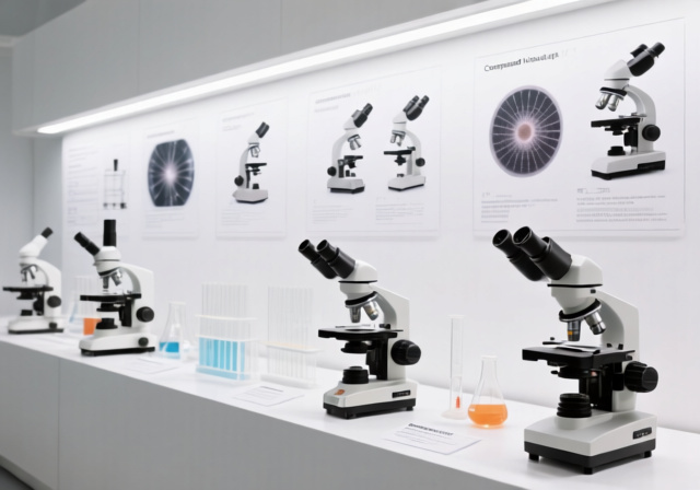
Compound light microscopes combine two lens systems. The objective lens sits close to the specimen and produces a magnified real image, which the eyepiece lens enlarges again for the viewer. Total magnification is the product of the two powers, so a 10x eyepiece paired with a 40x objective produces 400x total magnification. This is the standard bench microscope found in classrooms, labs, and clinics around the world.
Resolution is the smallest distance between two points that can still be told apart. It is not the same as magnification. A 1000x scope with poor optics will simply produce a larger, blurrier image. Ernst Abbe showed that the resolution of a light microscope is limited by the wavelength of visible light, which is why no conventional optical microscope can resolve objects smaller than about 200 nanometers, and why the practical resolution floor for most student scopes sits closer to 500 nanometers.
Electron microscopes sidestep that limit by using electron beams, whose effective wavelength can be thousands of times shorter than visible light. The trade-off is that specimens must be prepared in vacuum and usually stained with heavy metals. Transmission electron microscopes fire electrons through ultra-thin samples, while scanning electron microscopes raster a beam across the surface to build a 3D-looking image.
Sample preparation is half the art of microscopy. Most biological specimens are nearly transparent under a standard light scope, so scientists use stains to add contrast. Hematoxylin and eosin paint tissues in pinks and purples, fluorescent dyes tag specific proteins, and heavy-metal stains such as uranyl acetate make cell structures pop under the electron beam. The 19th-century discovery of synthetic dyes, championed by Paul Ehrlich, turned microscopy from a black-and-white curiosity into a full-color biological tool.
The unifying principle is elegant: by manipulating waves and contrast, microscopes turn invisible structure into a picture the human brain can interpret. That picture has, over and over, changed what we know about life, matter, and the universe.
Microscope design has branched into a dozen specialties, each tuned to a different kind of specimen or a different kind of question. The table below covers the ten most common types you will encounter, from the student bench scope to the atomic-resolution probe.
| Microscope Type | Magnification Range | Best For | Key Features |
|---|---|---|---|
| Compound Light | 40x – 1000x | Biological samples, classroom work, cells | Multiple lenses, affordable, stained specimens |
| Stereo (Dissecting) | 7x – 50x | 3D objects, insects, circuit boards | Two eyepieces, low magnification, 3D view |
| Digital | 20x – 200x | Classroom teaching, screen sharing, documentation | USB camera, live display on monitor |
| Phase Contrast | 100x – 1000x | Live, unstained cells in culture | Converts phase shifts to brightness, no stain needed |
| Dark Field | 100x – 1000x | Bacteria, flagella, spirochetes | Bright specimen on a black background |
| Fluorescence | 400x – 1000x | Tagged proteins, live-cell imaging, neuroscience | Excites fluorescent dyes or GFP-tagged molecules |
| Confocal | 400x – 2000x | Thick samples, 3D reconstructions of cells | Laser scanning, optical sectioning, crisp 3D stacks |
| Polarized Light | 40x – 600x | Geology, crystals, minerals, fibers | Uses crossed polarizers to reveal birefringence |
| Transmission Electron (TEM) | 1,000x – 10,000,000x | Virus structure, organelle ultrastructure | Electron beam passes through thin slice |
| Scanning Electron (SEM) | 20x – 1,000,000x | Surface details, 3D imaging of specimens | Raster beam, dramatic depth of field |
| Atomic Force (AFM) | Up to 1,000,000,000x (atomic) | Single atoms, biomolecules, materials surfaces | Mechanical tip “feels” the surface, no lens needed |
Readers curious about the foundational split in microscope design can explore the differences between simple and compound microscopes in more depth, including how a Leeuwenhoek-style single lens compares to a modern multi-lens bench scope. For electronics work, where surface detail and soldering joints matter more than cellular structure, the specialized microscope applications used in electronics repair tend to be stereo or digital models with long working distances.
Microscopes have moved well beyond the biology classroom. Today they shape fields from vaccine design to art conservation. Here are ten of the most striking ways microscopes are used right now:
Medical Diagnostics: Pathologists examine tissue biopsies to identify cancer cells, infections, and blood disorders. Roughly 80% of diagnostic decisions rely on some form of laboratory microscopy.
Drug Development: Pharmaceutical researchers use cryo-electron microscopy (a method that earned the 2017 Nobel Prize) to see how drug candidates bind to proteins, dramatically speeding the design of new medicines.
Forensic Science: Crime labs compare hair, fibers, gunshot residue, and trace evidence under stereoscopes and comparison microscopes that can split a single view into two samples side by side.
Environmental Monitoring: Microbiologists track algae blooms, waterborne parasites, and soil health using fluorescence and phase contrast scopes.
Materials Science and Nanotechnology: Electron and atomic force microscopes reveal the grain structure of metals, the arrangement of atoms in graphene, and the surface of engineered nanomaterials.
Electronics and Semiconductor Inspection: Chip fabs depend on optical and electron microscopes to inspect features only a few nanometers wide. Specialized microscope applications in electronics repair have also become essential on smaller bench scales.
Agricultural Research: Plant pathologists identify fungi, bacteria, and viruses that threaten crops, helping breeders develop resistant varieties.
Veterinary Medicine: Vets diagnose parasites, blood disorders, and skin conditions in pets and livestock with the same staining and microscopy techniques used in human medicine.
Education and Citizen Science: From elementary classrooms to community lab spaces, affordable digital microscopes let students and hobbyists see the hidden structure of everyday life. Pond water and bread mold remain the most popular starter specimens for home users.
Art and Archaeology Conservation: Conservators use microscopes to identify pigments, check the condition of ancient pigments, and document restoration work on paintings and manuscripts.
One of the most mind-blowing modern uses is in vaccine and pandemic research. Cryo-EM was instrumental in resolving the spike protein of SARS-CoV-2, which accelerated the development of mRNA vaccines. In a single decade, microscopy has gone from a slow, indirect structural technique to a real-time window onto the molecular machines of disease.
Whether you are a student using a school microscope for the first time or a researcher maintaining a multi-thousand-dollar instrument, a few habits keep your scope in good shape and your images in focus. The five rules below are the foundation of careful microscopy.
Start at the Lowest Magnification: Begin with the 4x objective to locate your specimen and center it in the field of view. Higher magnifications have smaller fields and shallower depth, making it easy to lose track of what you are looking at.
Set the Lighting Before You Focus the Lenses: Adjust the diaphragm and light intensity so the field is bright and even, but not glaring. Proper illumination reduces eye strain and is essential for contrast at higher powers.
Use the Coarse Focus Only at Low Power: The coarse focus knob moves the stage quickly and can slam a slide into a 100x objective. Switch to fine focus the moment you are at 10x or higher, and refocus carefully with each change of objective.
Keep Lenses Clean and Dry: Wipe optics only with proper lens tissue and a recommended cleaning solution. Fingerprints and dust scatter light and create hazy images, and scratching a coated lens can ruin it permanently.
Carry, Cover, and Store Carefully: Hold the scope with one hand on the arm and one under the base, always replace the dust cover, and store the instrument in a dry cabinet. For educators and parents choosing microscopes for educational purposes, these habits are the difference between a scope that lasts a decade and one that survives a school year.
Microscopy has always been a frontier science, and the last fifteen years have brought some of the most dramatic advances since Leeuwenhoek first sketched his “animalcules.” Two Nobel Prizes in particular mark the modern era. In 2014, Eric Betzig, Stefan Hell, and William Moerner won the Nobel Prize in Chemistry for super-resolution microscopy, the family of techniques (STED, PALM, and STORM) that finally broke the diffraction limit of light and made it possible to image single molecules inside living cells.
Three years later, in 2017, Jacques Dubochet, Joachim Frank, and Richard Henderson shared the Nobel Prize in Chemistry for developing cryo-electron microscopy. Cryo-EM flash-freezes samples in vitreous ice, allowing biologists to image proteins and viruses in their native states. The technique has transformed structural biology, drug discovery, and our understanding of diseases ranging from Alzheimer’s to Zika.
Artificial intelligence is the latest revolution. Modern microscopes generate terabytes of imaging data per day, far more than a human can analyze by eye. Convolutional neural networks now classify cells, track cancer progression, and even suggest which experiments a scientist should run next. AI-assisted microscopy has turned the instrument from a passive observer into a partner in discovery, helping researchers spot patterns that would have taken a lifetime to find manually.
Digital microscopy has also democratized the field. Affordable USB scopes connect to phones and laptops, putting magnification in the hands of hobbyists, citizen scientists, and students across the world. Combined with cloud storage and remote collaboration tools, today’s microscopes can be operated from across the planet, opening up global classrooms and shared research programs in ways that were unimaginable a generation ago.
From the first rock-crystal lens of antiquity to the AI-driven cryo-EM of 2026, the microscope has always done the same job: it shrinks the gap between human vision and the world that exists just past it. As long as there are smaller things to see, there will be a new generation of scientists finding new ways to see them.
The word microscope was coined in 1625 by Giovanni Faber, a member of the Accademia dei Lincei in Italy. He paired the Greek mikros (small) with skopein (to look at) to make the Latin microscopium, giving the instrument a name that has lasted four centuries.
There is no single answer, because microscopy has so many parents. Antonie van Leeuwenhoek is often called the father of microbiology for his single-lens scopes, while Robert Hooke is honored as the father of cell biology for the illustrations in Micrographia. Marcello Malpighi is sometimes called the father of microscopic anatomy for his work on capillaries and tissues.
The name comes from the Greek mikros, meaning small, and skopein, meaning to look at. Italian scholar Giovanni Faber coined the Latin form microscopium in 1625 when he proposed both microscope and telescope as a matched pair of names for the new lens instruments of the seventeenth century.
Even the most powerful optical microscope cannot resolve anything smaller than about 200 nanometers, and most student scopes bottom out around 500 nanometers. To see individual atoms you need a scanning tunneling microscope or an atomic force microscope, and to see inside living cells at the molecular level you need a transmission electron microscope or a cryo-EM.
Modern microscopes are used in medical diagnostics, drug development, forensic science, environmental monitoring, materials science, electronics inspection, agricultural research, veterinary medicine, classroom education, and art and archaeology conservation. Newer applications include cryo-EM for vaccine research and AI-assisted imaging of large biological datasets.
Yes, but only with the right setup. A standard bright-field student microscope at 400x or 1000x can show larger bacteria such as Bacillus or Staphylococcus when the slide is stained with a dye like methylene blue. Tiny bacteria such as the cholera vibrio often need a dark-field or fluorescence microscope to be seen clearly, which is why Robert Koch’s staining breakthroughs were so important in the 1880s.

![Best USB Microscope [cy]: 12 Top Models for Every Budget](https://revellphotography.com/wp-content/uploads/2026/09/featured-update-1123-1789497022706.jpg)