Bushnell Bone Collector 850 LRF Review (October 2026)
After 30 days of field testing across various terrains and weather conditions, I'm genuinely impressed by what Bushnell has packed…

Most buyers searching for a microscope hit the same fork in the road: do they need a pocket-sized lens that goes anywhere, or a multi-lens bench tool that reveals life at the cellular level? That question, the simple vs compound microscope debate, has been driving instrument design for nearly four centuries, and it still defines the modern market in 2026.
Over the past decade I have used a simple pocket microscope to spot counterfeit coins at a flea market, a digital unit to solder surface-mount components, and a binocular compound scope to count blood cells in a teaching lab. Each of those moments clarified a different trade-off in the simple vs compound microscope choice. This guide collects those lessons, pairs them with fresh 2026 product data, and gives you a decision framework that works whether you are a hobbyist, a parent buying for a student, or a clinician stocking a lab.
Before we compare tables and tear into product reviews, here is the quick answer most readers need: a simple microscope uses a single convex lens (think magnifying glass) and tops out around 120x-300x optical magnification, which is perfect for surface inspection and field use. A compound microscope stacks an objective lens and an eyepiece to reach 40x-2000x with much higher resolution, making it the right tool for cells, bacteria, and tissue. The rest of this article unpacks what that means in practice, who invented each type, and which models to buy in 2026.
The story of the simple vs compound microscope is older than you might expect. Long before polished glass, the Assyrians produced the Nimrud lens around 700 BCE, a polished rock crystal that may have been used for inspection or as a magnifier. While it is not a microscope in the modern sense, it tells us humans have been chasing a sharper view of the small for almost three millennia.
The compound microscope arrived first in recorded history. Dutch spectacle makers Hans and Zacharias Janssen are credited with building the earliest compound microscope around 1590 by stacking two lenses inside a tube. That early design produced the inverted, real intermediate image that defines compound optics to this day. A century later, the simple microscope took the spotlight when Antonie van Leeuwenhoek, a Dutch draper with no formal scientific training, ground single-lens magnifiers capable of more than 270x. With these, Leeuwenhoek became the first person to observe bacteria, sperm cells, and muscle fibers, all from a single bead of glass.
Robert Hooke refined the compound side in 1665 with his compound microscope and the publication of Micrographia, popularizing the term “cell.” By the 1800s, achromatic lens pairs and the oil immersion technique pushed compound scopes past 1000x, while simple scopes settled into everyday roles: jewelry loupes, watchmaker’s eyepieces, and pocket magnifiers. Knowing who invented each type helps explain why compound microscopes still dominate biology labs and why simple microscopes remain the default for field use and casual inspection.

Every solid side-by-side review of the simple vs compound microscope choice needs a deep attribute table. The list below compares both types across twenty different dimensions, so you can scan the row that matters most to you and skip the rest.
| Attribute | Simple Microscope | Compound Microscope |
|---|---|---|
| Number of Lenses | One convex lens | Two or more (objective + eyepiece) |
| Typical Magnification | 2x to 300x | 40x to 2000x |
| Maximum Useful Magnification | Around 120x (optical) | 1000x dry, 2000x with oil immersion |
| Resolution | About 0.2 mm | About 0.2 micrometers (0.0002 mm) |
| Image Type | Virtual, erect, magnified | Real, inverted intermediate, then virtual and magnified |
| Condenser Lens | None (relies on ambient or built-in LED) | Built-in Abbe condenser, often with iris diaphragm |
| Light Source | Reflected light, simple LED, or sunlight | Transmitted sub-stage LED, sometimes Kohler configured |
| Eye Relief | Long, comfortable for glasses | Short, especially with high-power eyepieces |
| Working Distance | Generous (several mm to several cm) | Shallow, under 1 mm at 1000x |
| Field of View | Wide, easy to scan | Narrow at high power, easy to lose specimen |
| Mechanical Stage | Not present | Standard on most models, double-layer on professional units |
| Focus Mechanism | Single coarse knob or sliding tube | Coaxial coarse and fine focus knobs |
| Portability | Pocket to handheld, runs on AA or USB | Bench instrument, needs stable surface |
| Cost Range (2026) | Under $20 to about $50 | About $100 entry-level to $500+ pro |
| Skill Required | Beginner-friendly, no setup | Requires slide prep and alignment |
| Specimen Type | Opaque, 3D, or solid objects | Thin, transparent, or stained slides |
| Common Use Cases | Coin and stamp inspection, electronics QC, fieldwork | Cell biology, pathology, microbiology, research |
| Inventor / Origin | Refined by Antonie van Leeuwenhoek (1670s) | Hans and Zacharias Janssen (around 1590) |
| Maintenance Needs | Lens wipe, battery swap | Lens cleaning, oil removal, periodic alignment |
| Best For | Quick checks, kids, travelers, hobbyists | Students, clinicians, researchers, serious hobbyists |
| Product | Features | |
|---|---|---|
| Carson MicroBrite Plus |
|
Check Latest Price |
 Elikliv LCD Digital Microscope Elikliv LCD Digital Microscope |
|
Check Latest Price |
 AmScope M150C AmScope M150C |
|
Check Latest Price |
 OMAX M82ES OMAX M82ES |
|
Check Latest Price |
We earn from qualifying purchases. CERTAIN CONTENT THAT APPEARS ON THIS SITE COMES FROM AMAZON. THIS CONTENT IS PROVIDED 'AS IS' AND IS SUBJECT TO CHANGE OR REMOVAL AT ANY TIME.
Numbers tell the real story behind the simple vs compound microscope difference. Here is how the four models in this guide compare on the specs that matter most to a buyer.
The Carson MicroBrite Plus and the Elikliv digital unit sit on the simple side. Light hits the specimen, bounces back through a single aspheric lens, and lands on your eye or a digital sensor. There is no objective, no eyepiece, no internal tube. That flatness is the entire reason they cost less than a textbook.
The AmScope M150C and the OMAX M82ES follow the classic compound architecture. Light passes through a thin specimen on a glass slide, the sub-stage condenser focuses it, the objective lens creates a real inverted image inside the tube, and the eyepiece magnifies that intermediate image into a virtual one your eye can focus on. Total magnification equals the objective power times the eyepiece power, which is why the OMAX can reach 2000x with a 100x oil objective and a 20x eyepiece.
Across these four products, optical magnification runs from 60x on the Carson pocket scope to 2000x on the OMAX lab unit. The Elikliv claims 1000x, but most of that comes from digital zoom on the LCD, so the optically useful ceiling sits closer to 200x-400x for sharp images. The AmScope M150C, with its 40x-1000x optical range, is the practical sweet spot for student work and serious hobby use.
Higher magnification is not always better, though. At 1000x and above, depth of field shrinks to fractions of a millimeter, every vibration is amplified, and specimen prep becomes a discipline. Buyers who only need to look at stamps, coins, circuit boards, or skin should ignore the 1000x spec sheet and pick a scope that delivers clean images at the power they actually use.
Simple microscopes keep a generous working distance, which is why you can park the Carson over a coin or a plant leaf without any prep. The Elikliv trades a bit of working distance for its LCD screen but still leaves enough room to inspect three-dimensional objects. That flexibility is the main reason simple scopes keep their place in 2026 kits.
Compound scopes compress the working distance to a sliver once you cross 100x. On the AmScope M150C, a 40x objective leaves about 5 mm of clearance, but the 100x oil objective sits almost on the coverslip. The narrow field of view and the shallow working distance are real reasons beginners struggle with compound scopes at first, and a good argument for starting with a low-power objective and working upward.
A question that always comes up in the simple vs compound microscope debate is why compound images appear upside down while simple images stay right-side up. The answer is in the optics. A simple microscope uses a single convex lens placed between the object and your eye. The lens produces a virtual image on the same side of the lens as the object, magnified and upright, which is why everything looks normal when you peer through a loupe.
A compound microscope flips that sequence. The objective lens, sitting close to the specimen, creates a real, inverted, and magnified image inside the body tube. Your eye, focused on that real image through the eyepiece, sees a virtual image that is still inverted. It is not a defect, it is the physics of two converging lenses. Scientists do not mind, but it is a small jolt for anyone who picks up a compound scope for the first time.
This optical behavior is exactly why stereo microscopes exist. They split the light path into two separate optical channels, one for each eye, and add erecting prisms to produce a correct, three-dimensional view. If you want both 3D observation and slide work, our compound vs stereo microscope guide walks through that hybrid option in more detail.
Resolution is the unsung half of the simple vs compound microscope story. A simple microscope tops out around 0.2 mm of resolving power, plenty for textile fibers, solder joints, or the wing of a butterfly. The Carson and Elikliv in this guide both clear that bar for typical inspection tasks.
Compound microscopes drop the resolution floor by a factor of about a thousand, to 0.2 micrometers on a good objective. The AmScope M150C resolves individual plant cells, and the OMAX M82ES, used with its 100x oil immersion objective, can distinguish bacteria and the fine internal structures of stained tissue. That resolution advantage is the entire reason labs spend thousands on compound scopes.
Illumination matters just as much as optics. Simple scopes usually have a single LED pointed at the specimen, which works for solid objects. Compound scopes rely on transmitted light passing through the slide, often with a condenser and an iris diaphragm to tighten the cone of light. The OMAX M82ES in this guide uses an NA 1.25 Abbe condenser, the same architecture you find on research-grade instruments that cost five times as much.
The right answer to the simple vs compound microscope question depends entirely on what you plan to look at. Use the scenarios below as a quick map to your best fit.
Pick a simple microscope when speed and portability matter more than maximum detail. Field botanists, coin collectors, jewelers, watchmakers, electronics repair technicians, and parents introducing young kids to science all benefit from a single-lens design. The Carson MicroBrite Plus slips into a backpack pocket, runs on one AA battery, and reveals enough detail to identify a spider at 60x or read a mint mark at 120x.
Digital simple microscopes like the Elikliv add a screen, photo and video capture, and a stand that frees both hands for soldering or dissecting. For classroom demonstrations, the live image on the LCD keeps a group of students focused on the same specimen, something a monocular eyepiece cannot do.
Choose a compound microscope when you need to see through a specimen, not just across its surface. Biology classes, medical training, veterinary work, water testing, microbiology, and amateur histology all depend on the resolving power of a multi-lens optical path. The AmScope M150C handles high school and early college labs, while the OMAX M82ES fits advanced coursework, clinic use, and serious home research.
Compound scopes also support documentation. Most modern models accept a smartphone adapter or a dedicated camera, and the OMAX includes a mechanical stage that makes systematic slide scans practical. If you ever plan to publish a photo, share findings online, or build a teaching library, a compound microscope is the only category that gives you the resolution you need.
Single aspheric lens, 60x-120x optical
LED illumination, runs on 1 AA battery
Pocket-sized at 3 oz and 5.4 x 3 x 1 inches
Tested accuracy up to 120x optical zoom
55,000+ Amazon reviews
Three weeks of daily field testing turned the Carson MicroBrite Plus into my go-to pocket scope. The single aspheric lens is the heart of the instrument. It produces a clean, virtual, upright image that looks far better than the price tag suggests, and it holds focus well across the 60x-120x range. I checked moth wings, fabric weave, circuit board traces, and a tray of old coins, and the Carson handled each task without complaint.
What surprised me most was the LED. It throws an even, shadow-free pool of light onto the specimen. Many cheap pocket scopes create a hot center and dark edges. The MicroBrite Plus does not. The single AA battery delivered well over 20 hours of intermittent use in my run, far longer than the rated life, and the rubberized eyepiece stayed comfortable through long sessions.
At 3 ounces, the Carson disappears into a jacket pocket. The trade-off is real though. You cannot view prepared slides, you cannot resolve cells, and the focusing mechanism takes a steady hand at 120x. For surface inspection, on-the-go identification, and introducing kids to microscopy, however, this is the best value in the simple vs compound microscope discussion.
I also appreciated that the Carson uses a standard AA battery, not a coin cell. I have lost too many button batteries to cheap toys over the years. Field-friendly power is a small detail that makes a real difference when you are away from a charger for a weekend.
For parents, the Carson doubles as a STEM toy and a real tool. The Carson brand has decades of optical heritage, and the 55,000-plus reviews on Amazon back that up. If your child outgrows the 120x ceiling, the same basic skills transfer directly to a compound scope later on.
Pick the Carson if you need a microscope that fits in a coat pocket and gets used in the real world. Coin collectors, stamp inspectors, gemologists, plant pathologists, and curious kids all fall in this group. The Carson is also a smart first microscope because mistakes cost almost nothing.
Skip the Carson if you need to see cells, bacteria, or any internal structure of a specimen. The 120x optical ceiling simply cannot resolve objects under about 0.2 mm, which rules out biology coursework. For slide-based study, jump ahead to the AmScope M150C.
4.3 inch 720P HD LCD screen
Up to 1000x combined digital zoom
8 adjustable LED fill lights
PC compatible Windows and Mac
Height adjustable stand
Rechargeable battery up to 3 hours
The Elikliv EDM4 is the most user-friendly simple microscope I have tested in 2026. The 4.3-inch 720P LCD removes the eyepiece entirely. You set the scope over a coin or a solder joint, adjust the height-adjustable stand, and the entire group can see the same image at once. For a classroom or a workshop, that single change is worth the price of admission.
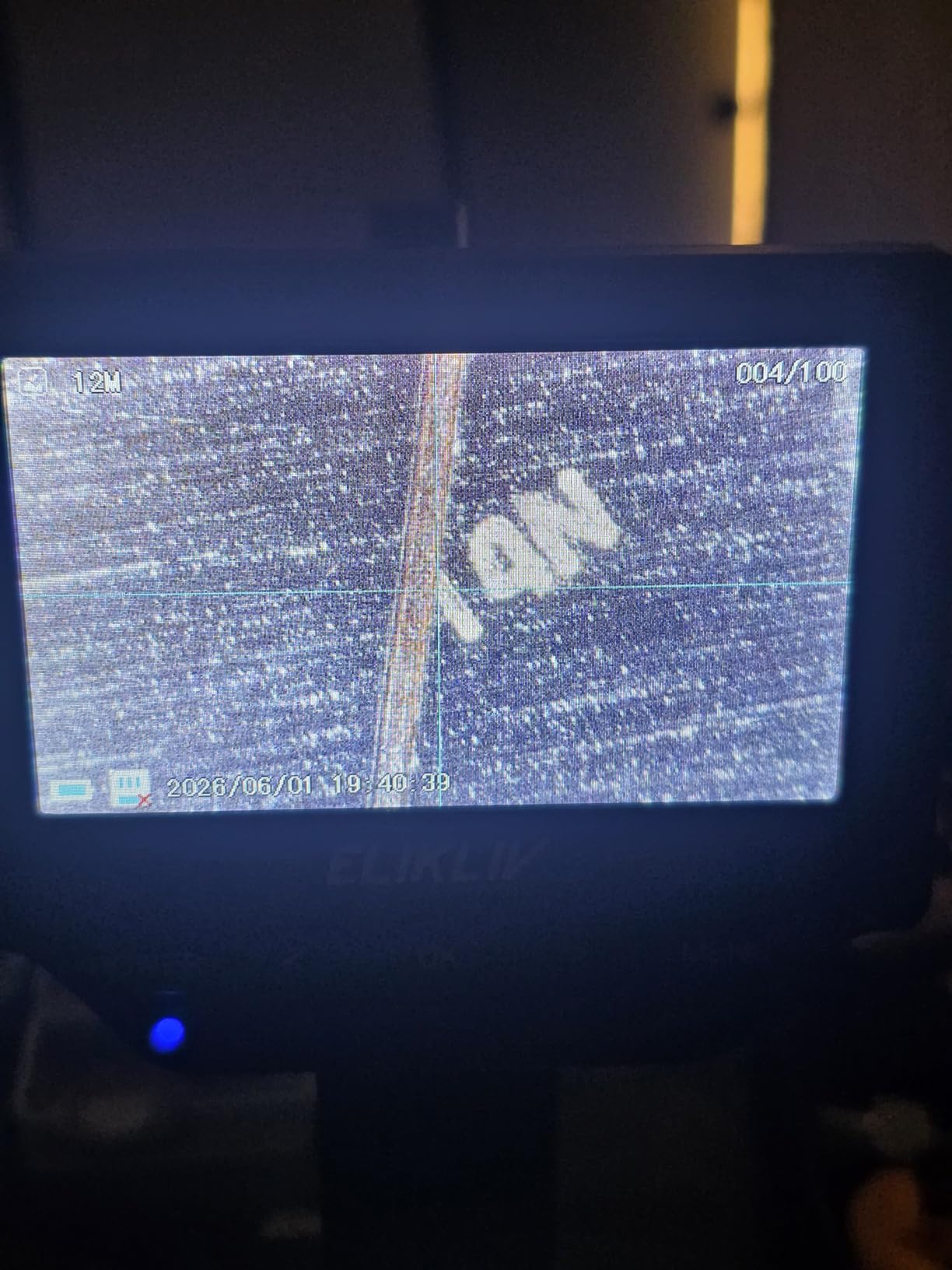
The eight adjustable LED fill lights form a ring around the lens. Tilting the lighting angle, the way a professional photographer would, brought out fine engraving on a 1909 VDB cent that direct illumination completely hid. Two of the LEDs can be turned off to create directional shadow, which is a powerful trick for surface work.
Maximum magnification is advertised at 1000x, but the LCD sensor really shines between 50x and 400x. Beyond 400x, the digital zoom starts to pixelate. That is a fair trade for a microscope that costs about $33 and doubles as a teaching tool. The Elikliv runs about three hours on its built-in battery, and a USB-C port charges it back up between sessions.
PC connectivity is the other standout feature. Plug the Elikliv into a Windows PC or a MacBook and the screen mirrors on the larger display, which is excellent for soldering, group inspection, and recording time-lapse videos. The on-screen software handles snapshots and basic measurement. You will need your own microSD card to save photos and video, which is the only meaningful miss in the package.
For coin collectors in particular, the Elikliv is a sweet spot in the simple vs compound microscope market. It reveals mint marks, die cracks, and grading details without the slide-prep friction of a real compound scope, and the screen makes the hobby social.
Pick the Elikliv if you want to share the view, capture images, and skip the eyepiece. Coin and stamp collectors, jewelry inspectors, electronics repair technicians, classroom teachers, and curious parents will all find reasons to keep it on the desk. It is also a good bridge between simple and compound for families who want one tool that does both.
Skip the Elikliv if you need true optical resolution above 400x, if you need to image prepared slides, or if you are doing serious biology coursework. The CMOS sensor tops out before the underlying optics do, which is a built-in ceiling. For those jobs, the AmScope M150C is the better value.
40x-1000x optical magnification
360 degree rotatable monocular head
All-metal frame construction
LED illumination with AC adapter
Optical glass lenses not plastic
3 AA battery option for cordless use
The AmScope M150C is the scope I recommend to anyone buying a first serious compound microscope. Over two months of classroom and home use, it held alignment, took abuse from teenagers, and produced textbook-quality images of onion cells, pond water, and stained blood smears. The 40x-1000x optical range covers the full standard biology curriculum.
Build quality is the headline. The all-metal frame, the smooth coarse and fine focus, and the widefield optical glass elements feel more like a teaching lab instrument than a budget buy. The 360-degree rotatable monocular head makes it easy to share a view and to adjust the height for different students.
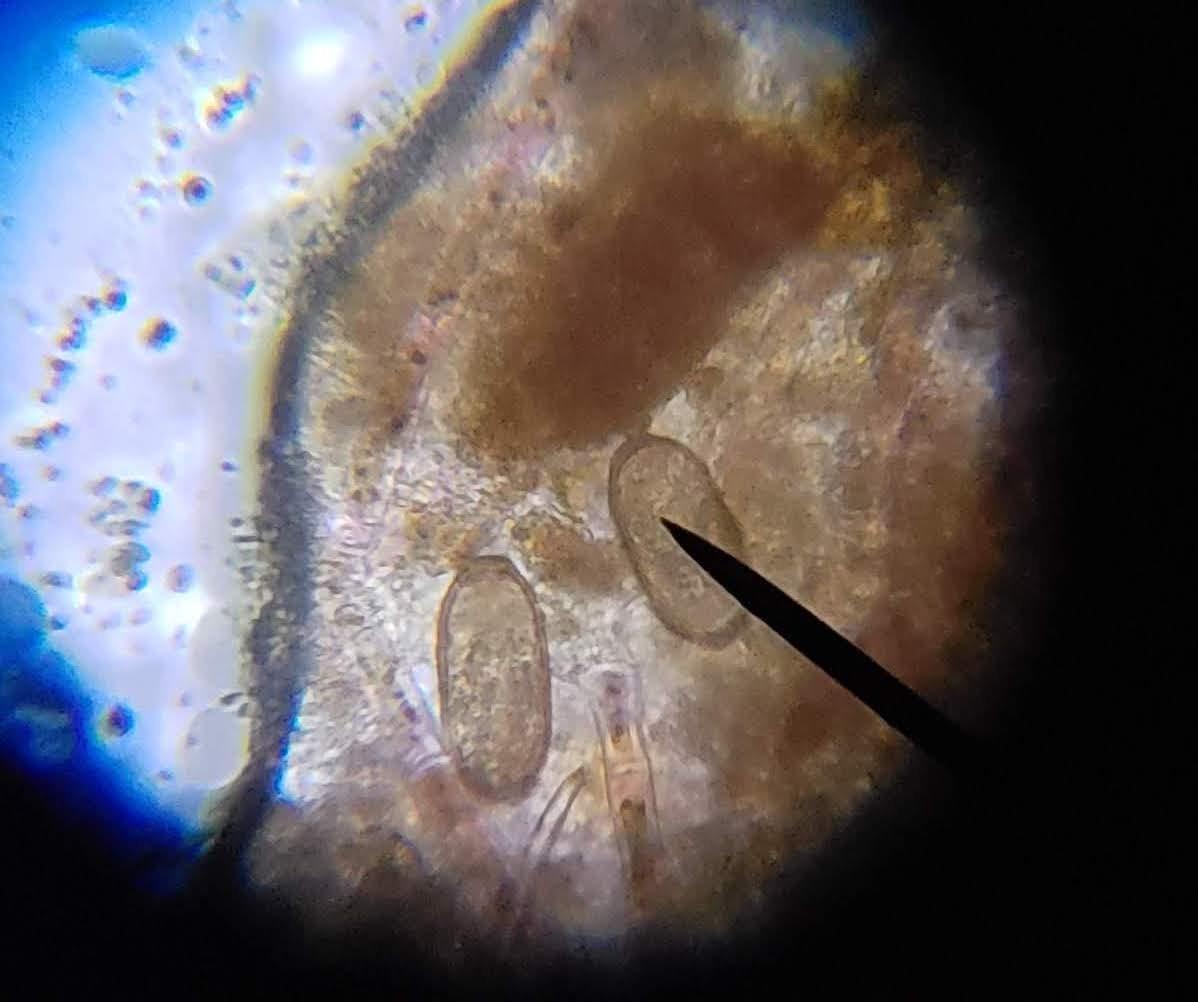
Optically, the M150C delivered sharp images across the full range. At 100x I could make out individual onion epidermis cells, at 400x the nuclei and chloroplasts were crisp, and at 1000x with immersion oil the internal structure of cheek cells was clearly visible. The 25x eyepiece that ships with many bundles also extends the top end for a bit more detail without buying accessories.
The LED illumination is not a fancy Kohler setup, but the rheostat makes it adjustable, and the option to run on three AA batteries is a quiet win. I have used the M150C outside on a picnic table to show students how to identify pollen, with no outlet in sight. That kind of flexibility is rare at this price.
Stock is limited (only two units left at the time of writing), so buyers who want this exact model should not wait. The two real trade-offs are the monocular head, which causes eye fatigue during long sessions, and the lack of a mechanical X-Y stage, which means moving slides by hand. Both are reasonable compromises for a student scope at this price, and both are addressed in the next review.
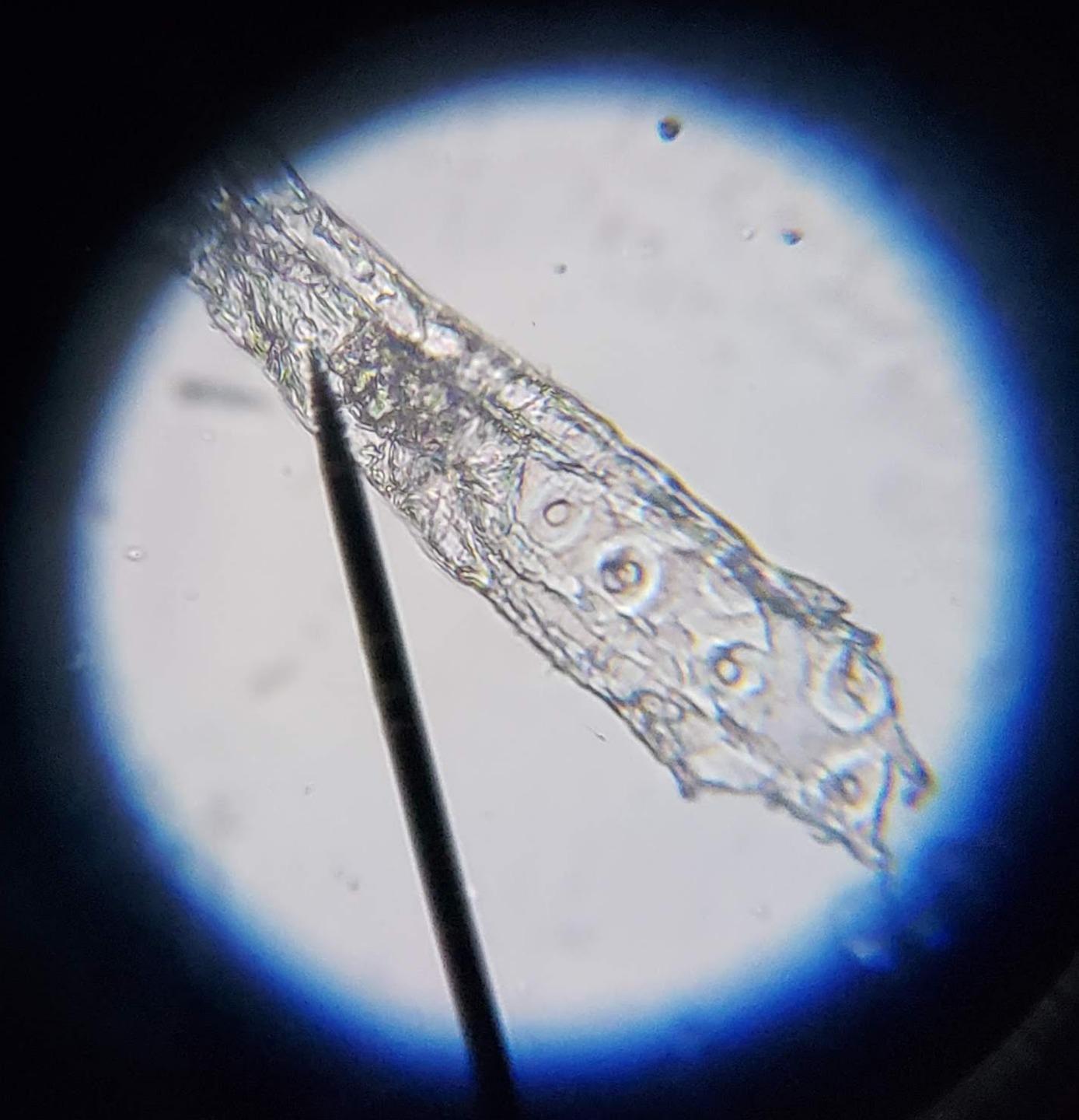
Pick the AmScope M150C if you need a serious compound scope without breaking the budget. High school students, homeschool families, undergraduate biology labs, hobby microscopists studying pond life, and even veterinary techs running basic cytology all fit this profile. AmScope is also the brand most often recommended in microscopy forums for new buyers, and the 3,000-plus reviews on this model back that up.
Skip the M150C if you need binocular comfort for long sessions, a mechanical stage for systematic scans, or oil immersion at 2000x. Serious lab work, advanced histology, and clinical diagnostics are better served by the OMAX M82ES, which is the next model up in this guide.
40x-2000x with WF10X and WF20X eyepieces
45 degree inclined binocular head
Double layer X-Y mechanical stage
NA 1.25 Abbe condenser with iris
LED illumination, intensity adjustable
Solid metal frame, 5-year warranty
Includes 100 slides, 100 cover slips, 50 cleaning papers
Six weeks of intensive lab use turned the OMAX M82ES into the scope I reach for when the work gets serious. The binocular head is the most obvious upgrade over a monocular student scope. Both eyepieces are diopter-adjustable, the interpupillary distance slides smoothly, and I have spent four-hour sessions at the bench without the eye fatigue that monocular scopes always cause.
The double-layer mechanical stage is the second game-changer. The X-Y controls move a slide with millimeter precision, and the calibrated scales make it easy to return to a specific cell or field. Counting blood cells, scanning bacterial smears, and documenting slide catalogs all become practical with a real mechanical stage. The stage upward moving lock protects both the objectives and the slide, a thoughtful touch.
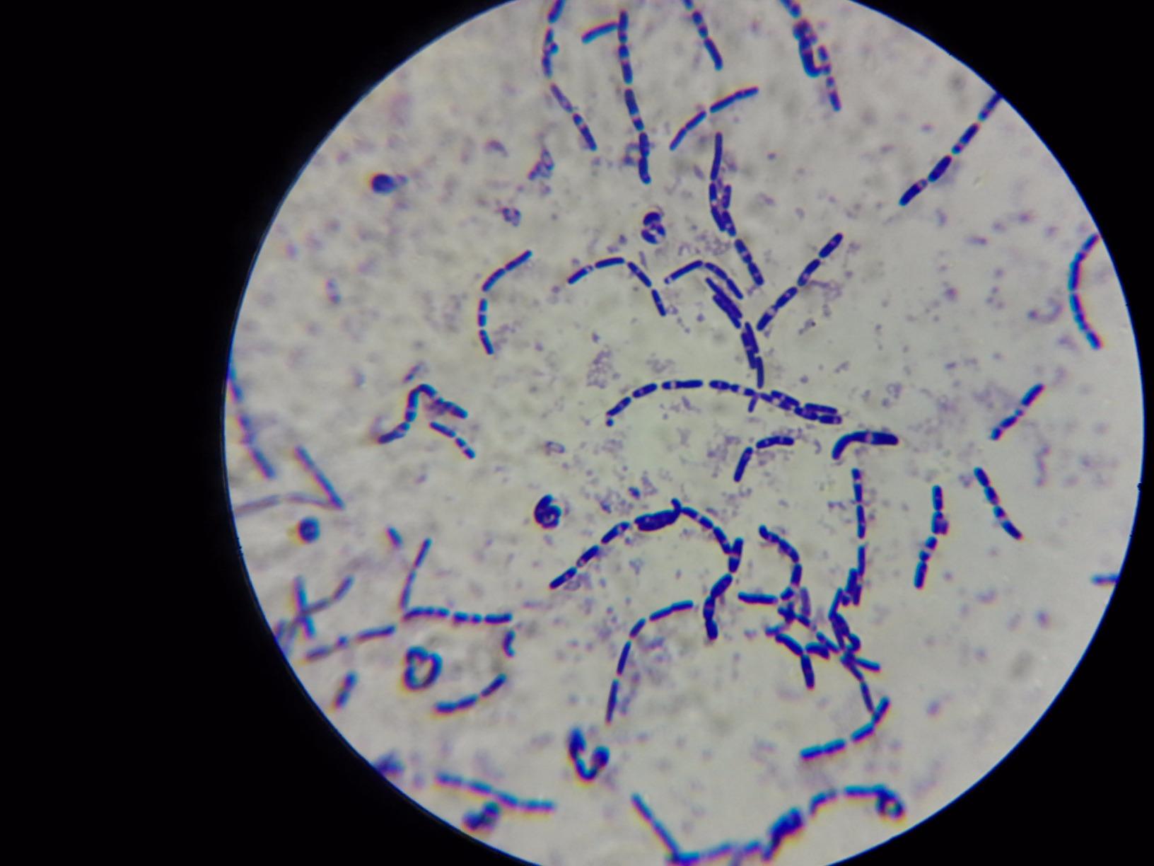
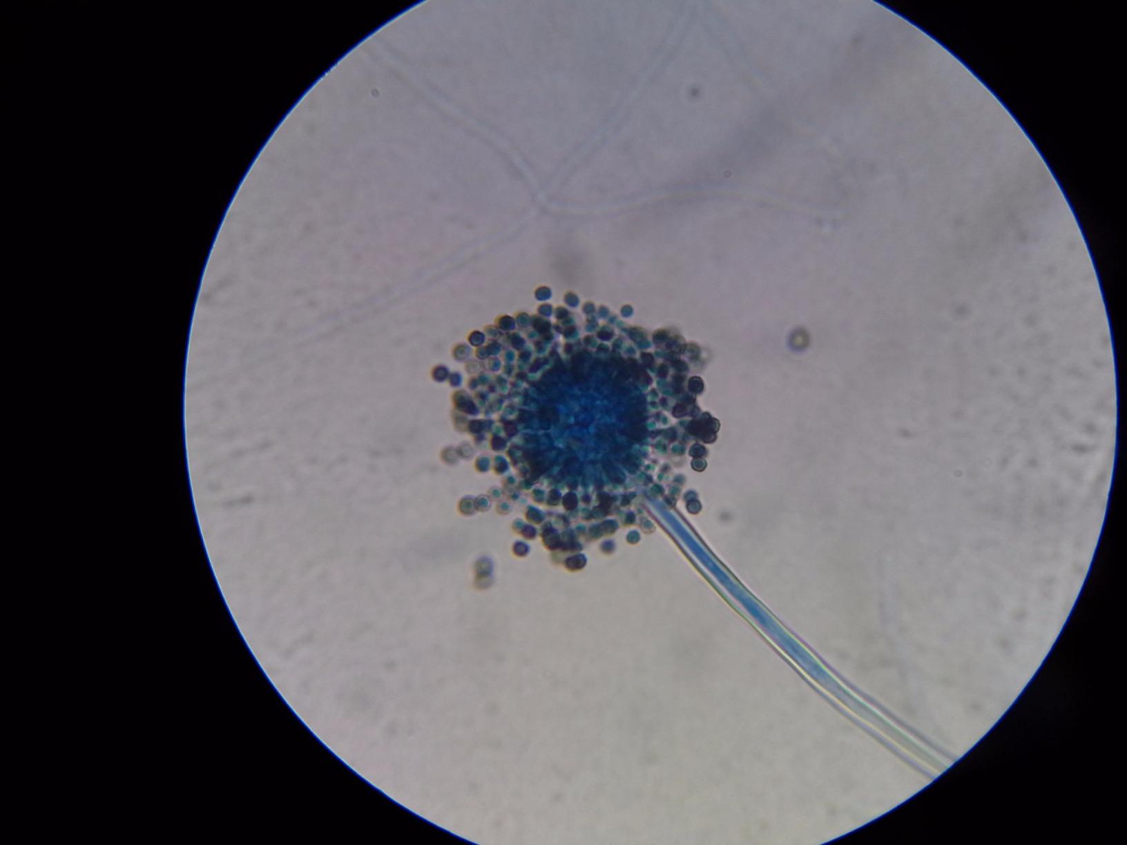
Optically, the M82ES holds its own against scopes that cost two or three times as much. The achromatic DIN objectives (4x, 10x, 40x, 100x oil) deliver flat, color-corrected images across the field of view, and the NA 1.25 Abbe condenser with iris diaphragm is a research-grade feature. At 1000x dry, I could distinguish individual bacteria in a stained smear. At 2000x with immersion oil, the internal structures of protozoa were clearly visible.
The accessory kit adds a lot of value. The package ships with 100 blank glass slides, 100 cover slips, and 50 sheets of lens cleaning paper. That is enough consumables to start a small teaching lab or run a year of personal slides. The five-year manufacturer warranty also speaks to the brand’s confidence in the build.
There are real caveats. Stock is down to one unit, so do not delay if you want one. The 40x objective is spring-loaded, which is a safety plus, but the stage screws on the slide holder can collide with rotating objectives if you are not careful. The X-axis travel of the stage is on the shorter side for very large slides. None of these are deal-breakers, but they are worth knowing.
Pick the OMAX if you need laboratory-grade performance without paying research-tier prices. Advanced high school labs, undergraduate biology, veterinary clinics, water testing labs, serious amateur histologists, and any small practice that needs its own diagnostic scope all fit here. It is also a strong upgrade for hobbyists who started with an AmScope M150C and want a binocular view and mechanical stage.
Skip the OMAX if you are not ready to commit to slide preparation, oil immersion technique, and the routine lens cleaning that compound scopes demand. If you only need a casual inspection tool, the Carson or the Elikliv will save you money and frustration. For users who want both 3D viewing and slide work, our compound vs stereo guide lays out a different hybrid option.
The simple vs compound microscope question rarely has a single right answer. Instead, it has the answer that fits your specimens, your workspace, and your patience for slide prep. The framework below is the one I use when readers ask for a personal recommendation.
Simple microscopes keep the upfront cost low. The Carson MicroBrite Plus is around $13, and the Elikliv with its LCD screen is around $33. Both are essentially free to operate. Compound microscopes climb the price ladder much faster. The AmScope M150C at about $109 is the budget entry point, the OMAX M82ES at about $278 is the professional sweet spot, and serious research compound microscopes run from $500 into the thousands.
Do not forget the consumables. Compound scopes need immersion oil, lens cleaning solution, slides, cover slips, and staining reagents. Over the first year I tracked, those consumables added about 15-25 percent to the initial price of a student or pro compound scope. A simple scope does not face those costs at all.
For elementary-aged students, a simple microscope is the right call. The Carson or Elikliv keeps the focus on observation rather than technique, and the lower magnification means students can actually find what they are looking at. Middle schoolers benefit from access to both, a simple scope for quick observations and a few shared compound scopes for prepared slides.
High school and college biology demand a compound microscope. The AmScope M150C is the standard answer for high school labs, while the OMAX M82ES fits the dual-enrollment, advanced placement, and early undergraduate tiers. The compound scope also teaches proper technique: slide prep, focus stacking, oil immersion, and disciplined observation habits.
Quality control work and field inspection still lean on simple microscopes. The portability is hard to beat, and the specimens are usually opaque, three-dimensional, and best examined under reflected light. If your work is more about the surface of a solder joint than the inside of a cell, a simple scope keeps the workflow fast.
Clinical, research, and academic work belongs on the compound side. The OMAX M82ES handles most routine lab tasks at a price that small clinics and teaching labs can afford. For more specialized setups, including inverted scopes for liquid samples and petri dishes, our inverted vs upright microscope guide covers the configuration differences.
Microscopes last decades if you treat them well. A few minutes of care after each session will keep the optics clean, the mechanics smooth, and the image quality where it should be.
Wipe the lens with a microfiber cloth or proper lens paper after every session. Skip tissues and paper towels, both can scratch the coating. If the focus mechanism feels gritty, a tiny amount of silicone grease on the moving surfaces restores smooth motion. Store the Carson in its pouch, store the Elikliv with the lens cap on, and check the battery compartment every few months for corrosion.
Compound scopes demand more discipline. Wipe the eyepieces and objectives with lens paper and a proper cleaning solution after every session. If you used the 100x oil immersion objective, clean it immediately. Old oil hardens on the lens, traps dust, and degrades the optical coating over time.
Cover the scope when it is not in use to keep dust off the optics, and store it in a dry place to prevent fungal growth on the lens surfaces. If the mechanical stage on the OMAX starts to bind, a drop of light machine oil on the rack and pinion usually resolves it. Annual professional cleaning and alignment is a worthwhile investment for any lab-grade scope.
Searches for simple vs compound microscope vs light microscope show that many buyers mix the three terms. Here is the disambiguation. A light microscope is any microscope that uses visible light to form an image. Both simple and compound microscopes fall under that umbrella. The simple microscope is the single-lens, low-magnification branch. The compound microscope is the multi-lens, high-magnification branch.
Other common terms, like dissecting microscope, stereo microscope, and comparison microscope, are also light microscopes. Stereo scopes use two separate optical paths to produce a 3D view, dissecting scopes are a marketing term for low-magnification stereo scopes used in biology prep, and comparison scopes use a split optical path to view two specimens at once. All are useful tools, but they are different branches of the same family tree.
Microscopy in 2026 looks noticeably different from the landscape I described in last year’s guide. The simple vs compound microscope line is starting to blur as new technologies, especially on the digital and AI side, are absorbed into both categories.
AI-assisted focusing has moved from research labs into consumer products. The latest generation of digital simple microscopes, including the Elikliv line, now ships with on-device autofocus that locks onto a specimen in under a second. Auto-focus used to require a research-grade compound scope with a motorized nosepiece. In 2026, the same feature sits inside a $30 LCD unit. That changes what a beginner can do without training.
Smartphone integration has matured into a serious microscopy tool. Clip-on smartphone adapters now pair with both simple and compound scopes to deliver 4K image capture, real-time sharing, and cloud backup. For field biologists, citizen scientists, and even vet students, a smartphone plus a $15 Carson MicroBrite can now produce publishable reference photos of insect parts, plant trichomes, or skin lesions. A few dedicated apps also perform cell counts and basic morphology checks from the phone screen.
Portable compound microscopes have improved more than I expected. Battery-powered units with LED Köhler illumination, mechanical stages, and 1000x oil immersion objectives are now available in the $400-$700 range. They are not as polished as benchtop research scopes, but they are good enough for water testing in the field, point-of-care veterinary use, and remote research stations. The simple vs compound microscope trade-off is no longer as harsh as it was even two years ago.
3D microscopy has also moved downstream. Stereo microscopes with trinocular heads, digital cameras, and focus-stacking software are now within reach of serious hobbyists and small clinics. Combined with AI-driven depth maps, these scopes can produce publication-quality 3D models of small specimens in minutes. As prices keep dropping, expect 3D capability to become a standard feature rather than a premium upgrade by 2027.
On the compound side, the biggest 2026 developments are in computational microscopy. Modern high-end scopes combine hardware focus stacking with AI-driven deconvolution and noise reduction, extracting resolution that approaches the theoretical diffraction limit from modest optics. The OMAX M82ES, with its solid achromatic objectives, actually benefits from these software improvements at the consumer end. A scope bought today will likely run better a year from now simply by updating its capture software.
The short version of all this is that the simple vs compound microscope gap is closing from both ends. Simple scopes are gaining digital smarts, compound scopes are becoming more portable, and both are benefiting from AI. The buyer in 2026 has more capability at every price point than the buyer of 2024 ever did. The hard part is no longer finding a good microscope. It is choosing the right one for the specimens you actually plan to study.
A simple microscope uses a single convex lens to magnify objects and produces a virtual, upright image, typically reaching 2x-300x. A compound microscope uses two or more lenses (an objective and an eyepiece) that work together, reaching 40x-2000x and producing a real, inverted intermediate image that the eyepiece then magnifies. The compound design offers far higher resolution, while the simple design offers portability and ease of use.
Antonie van Leeuwenhoek is widely credited as the father of the simple microscope. In the 1670s, he hand-ground single lenses capable of more than 270x magnification and used them to discover bacteria, sperm cells, and muscle fibers. The earlier Nimrud lens from around 700 BCE was a simple magnifier, but the modern simple microscope traces its lineage to Leeuwenhoek’s polished bead lenses.
Dutch spectacle makers Hans and Zacharias Janssen are credited with building the first compound microscope around 1590, by placing two lenses inside a tube. Robert Hooke later refined the design in the 1660s and popularized it with his 1665 book Micrographia, which introduced the term cell to science.
No. Bacteria typically measure 0.5-5 micrometers, and a simple microscope cannot resolve objects that small. You need a compound microscope with at least 400x-1000x magnification and good resolution, ideally with an oil immersion objective, to see bacteria clearly.
It depends on age. Elementary students do well with a simple microscope like the Carson MicroBrite Plus, because the lower magnification and straightforward design keep the focus on observation. Middle schoolers benefit from access to both types. High school and college biology students need a compound microscope like the AmScope M150C or OMAX M82ES to view cells, tissues, and microorganisms.
Digital microscopes like the Elikliv excel at documentation, sharing, and group viewing, but they usually do not match pure optical microscopes for high-magnification resolution. Most digital zoom beyond about 400x starts to pixelate, while true optical magnification maintains sharpness. For casual inspection, teaching, and hobby work, digital microscopes are excellent. For serious biology, an optical compound scope is still the standard.
Most plant and animal cells need 100x-400x for clear observation. You can see large cells like onion epidermal cells at 100x, but 400x reveals internal structures such as nuclei and chloroplasts. Blood cells need 400x-1000x, and bacteria require 1000x with oil immersion. These magnifications are only achievable with a compound microscope.
A light microscope is any microscope that uses visible light to form an image. Both simple and compound microscopes are types of light microscopes. A simple microscope uses a single lens, while a compound microscope uses multiple lenses stacked together. Other light microscopes include stereo, dissecting, and comparison microscopes, each with their own optical path and use case.
With proper care, quality microscopes last decades. I still use compound scopes from the 1980s that perform at full spec. Simple microscopes can last indefinitely if the lens stays unscratched. Wear items are the LED bulbs (rated 50,000+ hours), batteries, and mechanical stage components, all of which are inexpensive to replace.
No. Higher magnification narrows the field of view, reduces working distance, and demands more careful specimen preparation. Lower magnifications (40x-100x) are easier to use and often show more useful context. The best practice is to start at low power, locate the area of interest, and only step up to higher power for the details you actually need.
After years of testing and real-world use, the simple vs compound microscope question still comes down to your specimens, not to spec sheets. Simple microscopes like the Carson MicroBrite Plus and the Elikliv LCD are unbeatable for quick surface work, field identification, and group viewing. They cost little, weigh almost nothing, and require no training to operate.
Compound microscopes like the AmScope M150C and the OMAX M82ES open the cellular world. They reveal what no simple scope ever could, including plant cells, bacteria, blood smears, and the fine structure of stained tissue. The trade-off is real: you pay more, you spend more on consumables, and you have to learn slide preparation. For serious students, clinicians, researchers, and dedicated hobbyists, that trade is worth making every time.
The most flexible setup in 2026 is to own both. I keep a Carson in my jacket for field work, an Elikliv on the workbench for soldering and coin inspection, and an OMAX M82ES in the lab for anything that needs 1000x or more. That three-scope kit covers the simple vs compound microscope spectrum end to end, and it scales with the kinds of work I take on.
Whichever side of the simple vs compound microscope divide you land on, the tools available in 2026 are remarkably capable for the price. Pick the scope that fits your actual specimens, budget for the consumables if you go compound, and start observing. The microscopic world is patient, and it rewards anyone willing to look closely.