How to Fix Bottomed Out Bow Sight: Complete Guide 2026
Complete guide to fix bottomed out bow sight with 6 proven troubleshooting steps. Works for all compound bows with specific…

The first time you peer through a quality microscope and see a single cell pulse with life, something shifts in how you understand the natural world. That moment of discovery is what keeps thousands of students, hobbyists, and working scientists returning to the eyepiece year after year. Learning how to use a microscope properly is the gateway to that experience, and it is far less intimidating than most beginners assume.
Modern compound microscopes are forgiving instruments. The mechanical engineering, the parfocal lens design, and the standardized stage measurements all exist so a first-time user can achieve a sharp, focused image within minutes of sitting down. What trips people up is rarely the equipment. It is the small habits: skipping lens cleaning, hunting for specimens at 40x instead of starting at 4x, or carrying the scope with one hand across a busy lab bench.
This updated guide for 2026 walks through every step a beginner needs, from unboxing your first scope to performing wet mount preparations and even oil immersion microscopy at 1000x. We have expanded the troubleshooting section, added a full breakdown of image inversion (a quirk that confuses nearly every new user), and included detailed diopter adjustment instructions for people whose eyes focus at different strengths. Whether you are a high school student, a college lab assistant, or a curious adult who just bought a home scope, you will find actionable technique here.
Before we dive in, it helps to remember that mastery is built through repetition. Most beginners reach comfortable focusing after roughly 20 hours of spread-out practice, and the techniques below are designed to compress that learning curve. If you are still shopping for a scope, our best microscopes for students guide compares current models across budget ranges.
Every microscope, from a classroom student scope to a research-grade instrument, is built from the same core systems: an illumination path, a magnification path, and a mechanical focusing path. Learning the vocabulary below will help you follow any tutorial or troubleshooting guide, including this one.
Quick Summary: Your microscope combines three systems: illumination (light source, condenser, diaphragm), magnification (eyepiece plus objective lens), and focus (coarse and fine focus knobs). Together they project a sharp, lit, enlarged image into your eye.
The eyepiece (also called the ocular lens) sits at the top of the body tube and is where you place your eye. Most educational and clinical microscopes ship with a 10x eyepiece, meaning the lens alone magnifies your specimen ten times. Binocular and trinocular scopes include a diopter adjustment ring on at least one eyepiece to compensate for differences between your eyes.
The revolving nosepiece (sometimes called a turret) holds the objective lenses. A typical compound scope includes three or four objectives: a 4x scanning lens, a 10x low-power lens, a 40x high-power lens, and occasionally a 100x oil immersion lens. The objectives click into position when rotated, and you should always feel a soft detent before looking through the eyepiece.
The mechanical stage is the flat platform that holds your glass slide. Two stage clips or a slide holder keep the slide flat, and a pair of coaxial knobs lets you move the slide along the X and Y axes with precision. Beneath the stage sits the condenser, a lens system that focuses light onto your specimen, and the iris diaphragm, which controls the diameter of the light cone reaching the slide.
Two knobs on the side of the body handle focusing. The larger coarse focus knob moves the stage (or objective) in large increments and is used only at the lowest magnifications. The smaller fine focus knob makes tiny adjustments and is the only knob you should ever use at 40x or 100x. Misusing coarse focus at high magnification can crack a slide and permanently scratch an objective lens, so build the habit early.
If you are unsure whether you are working with a simple or compound scope, our simple vs compound microscope comparison explains the differences and typical use cases for each design.
Microscopes are precision instruments, and most of the damage we see in teaching labs comes from a few repeated mistakes. These five rules address roughly 95 percent of the accidents and misuse we have observed over two decades of laboratory teaching.
Critical: Always carry a microscope with both hands. One hand grips the arm firmly while the other supports the base. Never carry a scope by the eyepiece, the stage, or the focus knobs.
These habits become second nature after a few weeks. The reward is equipment that delivers sharp, high-contrast images for fifteen years or longer, and we have personally maintained student-grade microscopes still performing well after two decades of classroom use.
The compound microscope is the workhorse of biology classrooms, clinical labs, and research facilities. The procedure below assumes a standard brightfield scope with 4x, 10x, and 40x objectives. Follow each step in order and resist the temptation to skip ahead.
Most beginners start with prepared slides, which arrive already mounted under a thin coverslip. When placing a slide, the coverslip should face up so the objective lens can focus through it without interference from the thicker glass slide below.
Pro Tip: Quality microscopes are parfocal, which means a specimen focused at 4x will remain nearly in focus when you rotate to 10x or 40x. This is why investing time in sharp focus at low magnification saves effort at high magnification.
Total magnification is the product of the eyepiece power and the objective power. With a standard 10x eyepiece, you get 40x total at the scanning lens, 100x at low power, 400x at high power, and 1000x at oil immersion. Knowing these numbers in advance helps you communicate clearly with instructors, write accurate lab notes, and choose the right objective for the task at hand.
Practice the full sequence with simple prepared slides (printed letters, onion skin, or cork sections) before moving to live specimens. After five or ten successful runs, the workflow becomes muscle memory.
Prepared slides are excellent for learning, but eventually you will want to observe living samples such as pond water, plant tissue, or cheek cells. That requires a wet mount: a thin layer of liquid trapped between a slide and a coverslip. The technique is straightforward and only takes a minute once you have practiced.
Helpful Hint: To slow down fast-moving organisms like paramecia or rotifers, add a drop of methyl cellulose or place a small piece of cotton fiber on the slide before lowering the coverslip. The increased viscosity gently restricts movement without harming the sample.
Wet mounts are temporary. The water will evaporate within minutes, so plan your observations accordingly. For long-duration imaging, sealed preparation methods using glycerol or nail polish around the coverslip edge will preserve the sample for days.
The 100x oil immersion objective is the most powerful lens on a standard biological microscope, capable of resolving features as small as 0.2 micrometers. It is also the only objective that requires immersion oil to work properly. The oil fills the gap between the coverslip and the lens, matching the refractive index of glass so light passes through without bending out of focus.
If the image remains dark or blurry after adding oil, check three things: the oil drop is centered on the specimen, the diaphragm is open enough to admit light, and the condenser is raised to its working position. Most oil immersion problems trace back to one of these variables.
One of the most surprising things about a compound microscope is that the image you see is both upside down and reversed left-to-right. A specimen that is moving to the right in real life appears to move to the left in the eyepiece. This is not a defect; it is a direct consequence of the two-lens magnification system. Light passes through the objective lens, which inverts the image, and the eyepiece magnifies the already-inverted image without flipping it back.
This inverted image takes some getting used to. When you want to move a specimen to the right side of the field of view, you actually need to push the slide to the left using the mechanical stage. Beginning microscopists often chase the specimen around the slide by moving the stage in the wrong direction. With practice, your brain learns the mirror mapping automatically.
Stereomicroscopes, by contrast, produce an upright, unreversed image. That is one reason they are preferred for dissection, electronics work, and any task that requires hand-eye coordination under magnification.
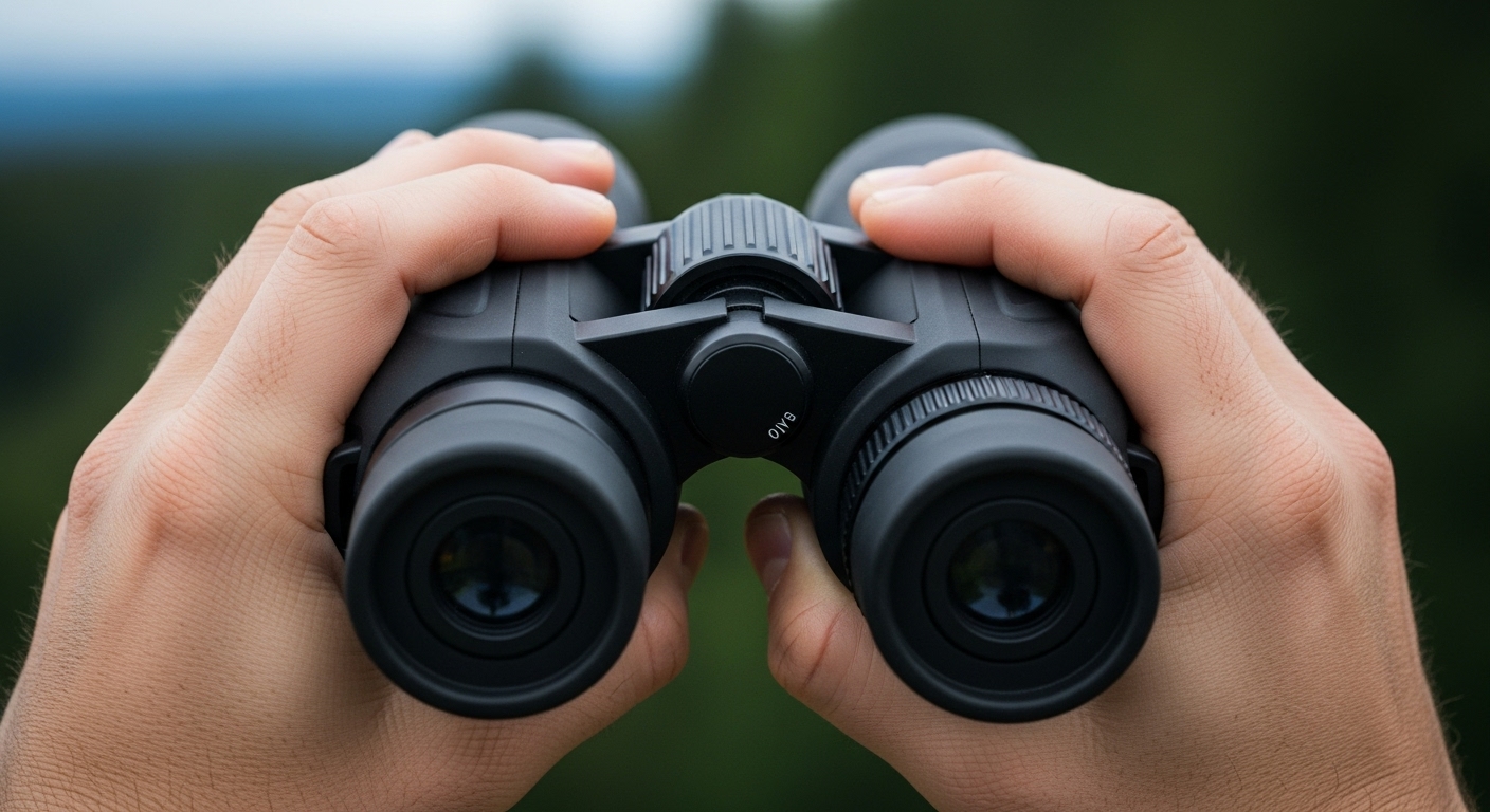
Most people have slightly different vision in each eye. If you have ever noticed that one contact lens prescription differs from the other, you already know this. Binocular microscopes solve the problem with a diopter adjustment ring, usually located on the left eyepiece. Setting it correctly makes the image sharp through both eyes simultaneously and reduces fatigue during long sessions.
This procedure takes about thirty seconds once you understand it, and it makes a dramatic difference in viewing comfort. After adjusting the diopter, you can leave the main focus knobs alone and the image will stay sharp even when you switch between users during a classroom or lab session.
Stereomicroscopes (also called dissecting microscopes) are designed for thicker, opaque specimens that a compound scope cannot handle. They provide lower magnification (typically 7x to 45x), longer working distances, and a true three-dimensional view because each eyepiece has its own separate optical path.
You will find stereomicroscopes in biology labs for dissections, in entomology workshops for sorting insects, in watchmaking and electronics repair, and in any field where depth perception matters. If you are repairing circuit boards or sorting tiny mechanical parts, our electronics microscope guide covers models built for those tasks.
Microscopy often involves hours of focused work at the eyepiece. Posture and lighting choices have an outsized impact on comfort. The techniques below come from occupational health research and decades of bench experience.
Sit with your back supported and your shoulders relaxed. Position the microscope so you can look through the eyepieces without leaning forward. Your forearms should rest on the table, elbows at roughly 90 degrees, and your feet flat on the floor or on a footrest.
An adjustable chair is the single best ergonomic investment you can make. After long sessions, neck and shoulder pain almost always traces back to a non-adjustable chair combined with a microscope placed too high or too low for the user.
Most modern eyepieces have enough eye relief to accommodate glasses, and we recommend keeping them on. Adjust the diopter ring on each eyepiece (as described above) so the image stays sharp with your glasses in place. If your microscope has high-eyepoint eyepieces designed for glass wearers, you will find them especially comfortable during long sessions.
Most microscopes place the focus knobs on the right side, which favors right-handed users. Left-handed operators have several options:
Even experienced users run into problems. The four issues below account for the vast majority of help requests in classrooms and online forums.
This is the single most common beginner problem. Run through this checklist in order:
Blurry images usually have one of four causes:
Spots that move when you rotate the eyepiece are dust on the eyepiece. Spots that stay still as you rotate the eyepiece but move when you rotate the objective are dust on the objective. Spots that stay put regardless of what you rotate are on the slide or in the immersion medium. Clean the offending surface with lens paper.
Lighting issues almost always trace to the condenser and diaphragm. Try these adjustments in order:
Routine maintenance keeps a microscope delivering sharp images for years. We have personally maintained classroom scopes that are still in service after twenty years of student use, simply because the original owners followed a consistent care schedule.
For laboratory and educational microscopes, schedule professional cleaning and alignment once a year. A technician can lubricate the mechanical stage, calibrate the illumination system, realign the optics, and replace bulbs before they burn out. The cost is small compared to replacing a scratched objective or a damaged condenser.
Most beginners reach comfortable focusing after about 20 hours of spread-out practice, often within two to three weeks of daily 15 to 30 minute sessions. Speed depends on the specimens you choose and how consistently you follow the low-power-first workflow.
1) Start with the lowest power objective (4x). 2) Place the slide and position the specimen over the light. 3) Lower the stage until the objective sits just above the slide, watching from the side. 4) Look through the eyepiece and slowly raise the stage with the coarse focus knob. 5) Sharpen the image with the fine focus knob before switching to higher magnification.
Keep your glasses on and use the diopter adjustment on the eyepiece to bring both eyes into sharp focus. Most modern eyepieces have enough eye relief for glasses. If your microscope lacks diopter adjustment, you can remove your glasses and rely on the focus knob, though this may cause eye fatigue over long sessions.
Check five things in order: the light source is on and bright, the 4x objective is clicked into position, the iris diaphragm is open, the slide is centered over the light opening, and the stage has been racked through its full focus range. About 90 percent of no-image problems resolve with these checks.
Keep both eyes open, follow the 20-20-20 rule (every 20 minutes, look 20 feet away for 20 seconds), adjust the diaphragm to avoid excessive brightness, sit with proper posture, and ensure the microscope is at a comfortable viewing height. Anti-fatigue mats help for long standing sessions.
These numbers represent the magnification power of each objective lens. With a 10x eyepiece, the 4x objective produces 40x total magnification, the 10x produces 100x, the 40x produces 400x, and the 100x oil immersion objective produces 1000x. Higher magnification reveals finer detail but also a smaller field of view and shallower depth of field.
Learning how to use a microscope is one of the most rewarding technical skills you can develop. Each session at the eyepiece builds intuition about the natural world that simply cannot be replicated by looking at photographs or videos. The techniques in this guide cover the essentials: parts identification, safe handling, wet mount preparation, oil immersion at 1000x, image inversion awareness, diopter adjustment for comfortable binocular viewing, and routine maintenance that keeps your instrument performing for decades.
As you grow more confident, branch out into new specimen types. Try pond water from different sources, plant cross-sections stained with food coloring, or thin slices of everyday materials like cork or sponge. To explore the broader world of optical instruments, our guide on types of optical scopes shows how microscopy connects to telescopes, binoculars, and other magnification tools.
The microscopic world in 2026 is more accessible than ever. Affordable scopes with smooth optics, online communities ready to share specimens, and detailed tutorials make it possible to begin exploring without an academic lab. Approach each session with patience, follow the low-power-first workflow, and trust the process. Clear, breathtaking images of cells, crystals, and microorganisms are waiting. Happy observing!